Label The Features Of An Animal Cell
Juapaving
Mar 25, 2025 · 7 min read
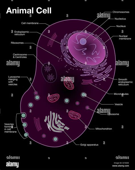
Table of Contents
Labeling the Features of an Animal Cell: A Comprehensive Guide
The animal cell, a fundamental unit of life, is a complex and fascinating structure teeming with activity. Understanding its components is crucial to grasping the intricacies of biology. This comprehensive guide delves into the features of an animal cell, providing detailed descriptions and clarifying their functions. We'll explore each organelle, highlighting its role in maintaining cellular health and overall organism function. By the end, you'll be well-equipped to accurately label and explain the purpose of each part of this remarkable cellular machine.
The Nucleus: The Control Center
The nucleus, often described as the "brain" of the cell, is the most prominent organelle. It's a large, membrane-bound structure that houses the cell's genetic material, DNA. This DNA is organized into structures called chromosomes, which contain the instructions for building and maintaining the entire organism.
Key Features within the Nucleus:
-
Nuclear Envelope: This double membrane surrounds the nucleus, regulating the passage of molecules in and out. It's punctuated by nuclear pores, which act as selective gateways, allowing specific molecules to enter and exit the nucleus.
-
Nucleolus: A dense region within the nucleus, the nucleolus is responsible for synthesizing ribosomal RNA (rRNA). These rRNA molecules are crucial components of ribosomes, the cellular machinery responsible for protein synthesis.
-
Chromatin: The uncondensed form of DNA, chromatin appears as a diffuse mass within the nucleus. During cell division, chromatin condenses into visible chromosomes.
Ribosomes: The Protein Factories
Ribosomes are the protein synthesis powerhouses of the cell. These small, granular organelles can be found free-floating in the cytoplasm or attached to the endoplasmic reticulum. They translate the genetic code from mRNA (messenger RNA) into proteins, the workhorses of the cell. Ribosomes are essential for virtually all cellular processes, from enzyme production to structural protein formation. The accurate construction of proteins is paramount to cell survival and functionality.
Endoplasmic Reticulum (ER): The Cellular Highway System
The endoplasmic reticulum (ER) is an extensive network of interconnected membranes extending throughout the cytoplasm. It exists in two forms:
Rough Endoplasmic Reticulum (RER):
The RER is studded with ribosomes, giving it its rough appearance. These ribosomes synthesize proteins destined for secretion or incorporation into cellular membranes. The RER plays a key role in protein folding, modification, and quality control. Improperly folded proteins are flagged for degradation, preventing errors that could have deleterious effects on the cell.
Smooth Endoplasmic Reticulum (SER):
The SER lacks ribosomes and is involved in various metabolic processes. Its functions include:
-
Lipid synthesis: The SER produces lipids, including phospholipids and steroids, crucial components of cell membranes.
-
Carbohydrate metabolism: The SER plays a role in glycogen breakdown (in liver cells) and the synthesis of certain carbohydrates.
-
Detoxification: In liver cells, the SER detoxifies harmful substances such as drugs and toxins. This detoxification process often involves modifying harmful substances so they are more easily excreted.
Golgi Apparatus: The Packaging and Shipping Center
The Golgi apparatus (or Golgi body) is a stack of flattened, membrane-bound sacs called cisternae. It receives proteins and lipids synthesized by the ER and further modifies, sorts, and packages them for transport to their final destinations – either within the cell or for secretion outside the cell. The Golgi apparatus is crucial for proper cellular organization and function, ensuring that molecules are delivered to the correct locations within or outside the cell. Dysfunction in the Golgi apparatus can lead to various cellular problems.
Mitochondria: The Powerhouses
Mitochondria are the "powerhouses" of the cell, generating most of the cell's energy in the form of ATP (adenosine triphosphate) through cellular respiration. This process involves the breakdown of glucose and other fuels to produce ATP, the cell's primary energy currency. Mitochondria have their own DNA and ribosomes, suggesting an endosymbiotic origin – they were once independent organisms that were engulfed by eukaryotic cells. Mitochondrial health is crucial for the overall function of the cell, and dysfunction can contribute to various diseases.
Lysosomes: The Recycling Centers
Lysosomes are membrane-bound organelles containing hydrolytic enzymes, which break down various cellular components, including worn-out organelles, cellular debris, and ingested materials. They maintain cellular cleanliness and prevent the accumulation of harmful substances. Lysosomes play a vital role in cellular homeostasis and waste management. Their proper functioning is crucial for preventing the buildup of potentially damaging materials.
Vacuoles: Storage and Waste Management
Vacuoles are membrane-bound sacs used for storage and waste management. They can store various substances, including water, nutrients, and waste products. In plant cells, a large central vacuole plays a crucial role in maintaining turgor pressure, keeping the cell firm. In animal cells, vacuoles are generally smaller and more numerous. They are involved in various processes, including endocytosis (engulfing materials from the outside) and exocytosis (releasing materials to the outside).
Cytoskeleton: The Cell's Internal Scaffolding
The cytoskeleton is a network of protein filaments that provides structural support and shape to the cell. It also plays a vital role in intracellular transport, cell division, and cell motility. The cytoskeleton is composed of three main types of filaments:
-
Microtubules: These are the largest filaments, providing structural support and acting as tracks for intracellular transport. They are also involved in cell division and cilia and flagella movement.
-
Microfilaments (Actin filaments): These are the smallest filaments, involved in cell shape, cell movement, and muscle contraction.
-
Intermediate filaments: These filaments provide mechanical strength and support to the cell.
Centrosomes: The Microtubule Organizing Centers
Centrosomes, located near the nucleus, are microtubule-organizing centers. They play a crucial role in cell division by organizing the mitotic spindle, which separates chromosomes during cell division. Each centrosome contains a pair of centrioles, cylindrical structures composed of microtubules.
Cell Membrane: The Protective Barrier
The cell membrane (or plasma membrane) is a selectively permeable barrier that encloses the cell's contents, separating the intracellular environment from the extracellular environment. It regulates the passage of molecules in and out of the cell, maintaining a stable internal environment. The cell membrane is composed of a phospholipid bilayer with embedded proteins. These proteins play various roles, including transport, cell signaling, and cell adhesion.
Peroxisomes: Detoxification and Lipid Metabolism
Peroxisomes are small, membrane-bound organelles that contain enzymes involved in various metabolic processes. They are involved in the breakdown of fatty acids and the detoxification of harmful substances. Peroxisomes produce hydrogen peroxide as a byproduct, which is then broken down by the enzyme catalase. Peroxisomes play essential roles in lipid metabolism and protecting the cell from oxidative damage.
Understanding the Interconnectedness of Organelles
It's crucial to understand that the organelles within an animal cell don't function in isolation. They are highly interconnected and work together in a coordinated manner to maintain cellular life. For example, the ER synthesizes proteins and lipids, which are then modified and packaged by the Golgi apparatus before being transported to their final destinations. Mitochondria provide the energy needed for these processes, and lysosomes help to recycle worn-out cellular components. The cytoskeleton provides the infrastructure for intracellular transport and cell division. This intricate interplay between organelles ensures the efficient and coordinated functioning of the cell.
Conclusion: Mastering Animal Cell Structure and Function
By understanding the individual features and interconnected functions of the organelles within an animal cell, we gain a profound appreciation for the complexity and beauty of life at the cellular level. This detailed guide provides a solid foundation for further exploration of cellular biology and its vast implications for understanding health, disease, and the fundamental processes of life itself. The ability to accurately label and describe the function of each organelle is a cornerstone of a deep understanding of this crucial biological unit. Remember to always approach the study of cell biology with curiosity and a desire to unravel the mysteries that lie within these microscopic worlds.
Latest Posts
Latest Posts
-
The Reaction Between An Organic Acid And An Alcohol Produces
Mar 28, 2025
-
Tap Water Mixture Or Pure Substance
Mar 28, 2025
-
The Role Of The Light Harvesting Complex Is To
Mar 28, 2025
-
Electrical Conductivity Physical Or Chemical Property
Mar 28, 2025
-
Why Do Atoms Want 8 Valence Electrons
Mar 28, 2025
Related Post
Thank you for visiting our website which covers about Label The Features Of An Animal Cell . We hope the information provided has been useful to you. Feel free to contact us if you have any questions or need further assistance. See you next time and don't miss to bookmark.
