Difference Between Plant Mitosis And Animal Mitosis
Juapaving
Mar 26, 2025 · 5 min read
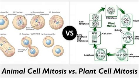
Table of Contents
Unveiling the Subtle Differences: Plant vs. Animal Mitosis
Cell division, a fundamental process in all living organisms, ensures growth, repair, and reproduction. Mitosis, a type of cell division, plays a crucial role in this process, creating two identical daughter cells from a single parent cell. While the fundamental mechanisms of mitosis are remarkably similar across eukaryotes (organisms with a membrane-bound nucleus), subtle yet significant differences exist between plant and animal mitosis, primarily stemming from the structural differences between plant and animal cells. Understanding these distinctions provides deeper insight into the cellular intricacies of life.
The Shared Fundamentals of Mitosis: A Quick Recap
Before delving into the specifics of plant versus animal mitosis, let's briefly review the common stages of mitosis:
Prophase:
- Chromosome Condensation: The replicated chromosomes, each consisting of two sister chromatids joined at the centromere, condense and become visible under a microscope.
- Nuclear Envelope Breakdown: The nuclear membrane surrounding the nucleus disintegrates, allowing the chromosomes to access the cytoplasm.
- Spindle Fiber Formation: Microtubules, the building blocks of the mitotic spindle, begin to assemble from centrosomes (in animal cells) or other microtubule organizing centers (MTOCs) in plant cells. These fibers will play a critical role in chromosome segregation.
Metaphase:
- Chromosome Alignment: The condensed chromosomes align along the metaphase plate, an imaginary plane equidistant from the two poles of the spindle. This precise alignment ensures equal distribution of chromosomes to the daughter cells.
- Spindle Fiber Attachment: Kinetochore microtubules, a subset of spindle fibers, attach to the kinetochores, protein complexes located at the centromeres of each chromosome. These attachments are crucial for chromosome movement.
Anaphase:
- Sister Chromatid Separation: The sister chromatids are pulled apart by the shortening of the kinetochore microtubules, moving towards opposite poles of the cell.
- Poleward Movement: Each separated chromatid, now considered an independent chromosome, travels towards the opposite pole, driven by the dynamic properties of the microtubules.
Telophase:
- Chromosome Decondensation: The chromosomes reach the poles and begin to decondense, returning to their less compact state.
- Nuclear Envelope Reformation: A new nuclear envelope forms around each set of chromosomes, creating two separate nuclei.
- Cytokinesis Initiation: The process of cytokinesis, the division of the cytoplasm, begins, leading to the formation of two distinct daughter cells.
Key Differences Between Plant and Animal Mitosis
While the general steps of mitosis are conserved, several crucial differences distinguish plant and animal mitosis:
1. Centrosomes and Spindle Formation:
- Animal Cells: Animal cells possess centrosomes, which are microtubule-organizing centers located near the nucleus. During prophase, centrosomes duplicate and migrate to opposite poles of the cell, forming the poles of the mitotic spindle. Microtubules radiate from the centrosomes, forming the spindle apparatus.
- Plant Cells: Plant cells lack clearly defined centrosomes. Instead, microtubule organizing centers are dispersed throughout the cytoplasm. The spindle apparatus forms without the distinct organization seen in animal cells. The spindle poles are defined by clusters of microtubules, rather than by centrosomes.
2. Cell Plate Formation vs. Cleavage Furrow:
- Animal Cells: Cytokinesis in animal cells involves the formation of a cleavage furrow. A contractile ring of actin filaments forms beneath the plasma membrane, constricting the cell from the outside inwards, eventually pinching the cell into two daughter cells.
- Plant Cells: Plant cells, surrounded by a rigid cell wall, cannot undergo cytokinesis via cleavage furrow formation. Instead, a cell plate forms in the center of the cell, gradually expanding outwards to partition the cytoplasm. The cell plate is constructed from vesicles derived from the Golgi apparatus, containing components for the new cell wall. This process results in the formation of a new cell wall between the two daughter cells.
3. Preprophase Band:
- Plant Cells: A unique feature of plant mitosis is the preprophase band. This is a transient band of microtubules that appears in the late G2 phase (the phase preceding mitosis) and encircles the nucleus. This band predicts the future site of the cell plate formation during cytokinesis, ensuring accurate cell division and maintaining the organized structure of plant tissues. This structure is absent in animal cell mitosis.
4. Cytokinesis Timing:
- Animal Cells: In animal cells, cytokinesis typically begins during anaphase and completes shortly after telophase. The timing is closely coordinated with the separation of chromosomes.
- Plant Cells: In plant cells, cytokinesis is a more protracted process. While the initiation overlaps with anaphase and telophase, the formation and expansion of the cell plate can take significantly longer.
5. Role of Cell Wall:
- Animal Cells: Animal cells lack a rigid cell wall, facilitating the flexibility required for cleavage furrow formation during cytokinesis.
- Plant Cells: The presence of a rigid cell wall dictates the different mechanism of cytokinesis in plant cells, requiring the construction of a new cell wall (the cell plate) to divide the cell. This is a fundamental difference that reflects the structural constraints imposed by the cell wall.
Detailed Comparison Table: Plant vs. Animal Mitosis
| Feature | Plant Mitosis | Animal Mitosis |
|---|---|---|
| Centrosomes | Absent; Microtubule Organizing Centers dispersed | Present; Two centrosomes forming spindle poles |
| Spindle Formation | Diffuse; No defined poles initially | From centrosomes; Well-defined spindle poles |
| Cytokinesis | Cell plate formation | Cleavage furrow formation |
| Preprophase Band | Present; Predicts cell plate formation | Absent |
| Cell Wall | Present; Influences cytokinesis | Absent |
| Cytokinesis Timing | Later, more protracted | Earlier, more rapid |
Evolutionary Significance of the Differences
The differences between plant and animal mitosis are not simply arbitrary variations. They reflect the evolutionary adaptations of these two distinct lineages to their respective environments and structural requirements. The rigid cell wall in plants necessitates the development of a unique mechanism for cytokinesis, the cell plate formation, unlike the flexible cytokinesis in animal cells. The absence of centrosomes in plants might represent a different strategy for organizing microtubules, possibly reflecting ancient evolutionary divergence. Further research continues to explore the deeper evolutionary implications of these differences.
Conclusion: A Tale of Two Mitoses
Plant and animal mitosis, while sharing the fundamental steps of chromosome segregation and cell division, exhibit distinct features reflecting their evolutionary histories and cellular structures. The contrasting mechanisms of cytokinesis, the presence or absence of centrosomes, and the unique preprophase band in plants highlight the diversity of cellular processes within the eukaryotic domain. Understanding these differences enhances our comprehension of the fundamental biological processes that underlie the growth, development, and reproduction of all life. Future research into the molecular mechanisms underlying these variations promises to unveil further insights into the fascinating world of cell biology and evolution.
Latest Posts
Latest Posts
-
Which Quadrilaterals Have Diagonals That Bisect Opposite Angles
Mar 29, 2025
-
List All The Factors Of 15
Mar 29, 2025
-
Is The Square Root Of 72 Rational Or Irrational
Mar 29, 2025
-
One Pair Of Opposite Sides Are Parallel
Mar 29, 2025
-
What Is The Relationship Between Density Mass And Volume
Mar 29, 2025
Related Post
Thank you for visiting our website which covers about Difference Between Plant Mitosis And Animal Mitosis . We hope the information provided has been useful to you. Feel free to contact us if you have any questions or need further assistance. See you next time and don't miss to bookmark.
