The Study Of The Tissues Is Called
Juapaving
Mar 25, 2025 · 6 min read
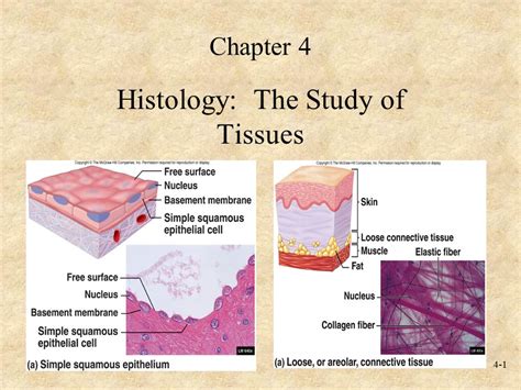
Table of Contents
- The Study Of The Tissues Is Called
- Table of Contents
- The Study of Tissues is Called Histology: A Deep Dive into the Microscopic World
- What is Histology?
- The Significance of Studying Tissues
- Histological Techniques: Preparing Tissues for Examination
- 1. Tissue Fixation: Preserving the Tissue Structure
- 2. Tissue Processing: Dehydration and Embedding
- 3. Sectioning: Creating Thin Tissue Slices
- 4. Staining: Visualizing Tissue Components
- 5. Microscopy: Visualization and Analysis
- Types of Tissues: An Overview
- 1. Epithelial Tissue: Covering and Lining
- 2. Connective Tissue: Support and Connection
- 3. Muscle Tissue: Movement
- 4. Nervous Tissue: Communication
- Applications of Histology
- The Future of Histology
- Latest Posts
- Latest Posts
- Related Post
The Study of Tissues is Called Histology: A Deep Dive into the Microscopic World
The study of tissues is called histology. Histology is a cornerstone of biology and medicine, providing crucial insights into the structure and function of living organisms. It bridges the gap between gross anatomy (the study of large body structures) and cell biology (the study of individual cells), offering a critical intermediate level of understanding. This detailed exploration will delve into the fascinating world of histology, covering its techniques, applications, and the importance of understanding tissue organization in health and disease.
What is Histology?
Histology, derived from the Greek words "histos" (tissue) and "logos" (study), is the microscopic study of the structure and composition of tissues. It involves examining thin sections of biological specimens using various microscopic techniques to identify different cell types, their organization, and the extracellular matrix that surrounds them. This detailed analysis reveals valuable information about tissue function, development, and pathology.
The Significance of Studying Tissues
Understanding tissue structure is paramount for several reasons:
-
Disease Diagnosis: Histopathological examination of tissue samples (biopsies) is a crucial diagnostic tool in medicine. Identifying abnormal tissue structures, such as cancerous cells or inflammatory infiltrates, allows for accurate diagnosis and treatment planning.
-
Understanding Physiological Processes: Histology illuminates the intricate relationships between cell structure and function. By observing the arrangement of cells and extracellular components, we can gain a deeper understanding of how tissues carry out their specific roles within the organism.
-
Developmental Biology: Histology plays a critical role in understanding the development of tissues and organs from embryonic stages. Analyzing tissue changes during development allows scientists to study how cells differentiate and organize into complex structures.
-
Research and Development: Histological techniques are instrumental in various research fields, including pharmacology, toxicology, and regenerative medicine. Researchers utilize these techniques to study the effects of drugs, toxins, and therapies on tissue structure and function.
Histological Techniques: Preparing Tissues for Examination
Preparing tissues for histological examination is a meticulous process involving several crucial steps:
1. Tissue Fixation: Preserving the Tissue Structure
Immediately after removal from the organism, tissues must be fixed to prevent degradation and preserve their structural integrity. Fixatives, such as formalin (formaldehyde), crosslink proteins and prevent enzymatic degradation, maintaining the tissue's original architecture. The choice of fixative depends on the type of tissue and the specific histological staining method to be used.
2. Tissue Processing: Dehydration and Embedding
Following fixation, tissues undergo a series of processing steps to prepare them for sectioning. This involves dehydration, using increasing concentrations of alcohol to remove water from the tissue, followed by clearing, using a solvent that is miscible with both alcohol and embedding medium. Finally, the tissue is embedded in a solid medium, such as paraffin wax or resin, providing support for sectioning.
3. Sectioning: Creating Thin Tissue Slices
The embedded tissue block is then sectioned using a microtome, a precision instrument that produces thin slices (typically 5-10 micrometers thick). These thin sections are crucial for allowing light to pass through the tissue during microscopy. The sections are then mounted onto glass slides for staining.
4. Staining: Visualizing Tissue Components
Staining techniques are essential for visualizing different components of the tissue. Hematoxylin and eosin (H&E) staining is the most common technique, where hematoxylin stains nuclei blue and eosin stains the cytoplasm and extracellular matrix pink. However, numerous other specialized stains exist, targeting specific cellular components or molecules, such as collagen, elastin, or specific enzymes. These specialized stains enhance the visualization of specific tissue structures and aid in accurate diagnosis. Immunohistochemistry (IHC) and in situ hybridization (ISH) are advanced techniques used to detect specific proteins and nucleic acids within tissue samples, respectively, which are very useful in diagnosing certain cancers.
5. Microscopy: Visualization and Analysis
Finally, the stained tissue sections are examined using a microscope. Light microscopy is commonly used for routine histological analysis, but electron microscopy (both transmission and scanning) provides higher resolution images, allowing visualization of subcellular structures. Digital microscopy and image analysis software are increasingly used for quantification and analysis of histological data.
Types of Tissues: An Overview
Animal tissues are broadly classified into four main types:
1. Epithelial Tissue: Covering and Lining
Epithelial tissue forms the coverings and linings of body surfaces, cavities, and organs. It is characterized by tightly packed cells with minimal extracellular matrix. Epithelial tissues are classified based on cell shape (squamous, cuboidal, columnar) and layering (simple, stratified, pseudostratified). Functions include protection, secretion, absorption, and excretion. Examples include the epidermis of the skin, lining of the digestive tract, and lining of the alveoli in the lungs.
Specialized Epithelial Tissues:
- Glandular epithelium: Specialized epithelial cells that secrete substances, such as hormones or mucus. Glands can be exocrine (secreting into ducts) or endocrine (secreting into the bloodstream).
- Sensory epithelium: Specialized epithelial cells that detect stimuli, such as light, sound, or taste.
2. Connective Tissue: Support and Connection
Connective tissue provides support, connects different tissues, and transports substances throughout the body. It's characterized by abundant extracellular matrix, which consists of ground substance and protein fibers (collagen, elastin, reticular). Different types of connective tissue include:
- Loose connective tissue: Provides support and cushions organs.
- Dense connective tissue: Provides strong support, found in tendons and ligaments.
- Cartilage: Provides flexible support, found in joints and ears.
- Bone: Provides rigid support and protection.
- Blood: A fluid connective tissue that transports oxygen, nutrients, and waste products.
- Adipose tissue: Stores energy in the form of fat.
3. Muscle Tissue: Movement
Muscle tissue is responsible for movement. There are three types of muscle tissue:
- Skeletal muscle: Attached to bones, responsible for voluntary movement. Characterized by striated appearance under the microscope.
- Smooth muscle: Found in the walls of internal organs, responsible for involuntary movement. Lacks striations.
- Cardiac muscle: Found only in the heart, responsible for pumping blood. Characterized by striations and intercalated discs.
4. Nervous Tissue: Communication
Nervous tissue transmits information throughout the body. It is composed of neurons (nerve cells) and glial cells (supporting cells). Neurons transmit electrical signals, while glial cells provide support and protection. The brain, spinal cord, and nerves are composed primarily of nervous tissue.
Applications of Histology
Histology is a vital tool in numerous fields:
-
Clinical Pathology: Histopathological examination of biopsies is crucial for diagnosing diseases, such as cancer, inflammatory conditions, and infectious diseases.
-
Forensic Pathology: Histology aids in determining the cause and manner of death in forensic investigations.
-
Veterinary Pathology: Similar to human medicine, histology is essential for diagnosing diseases in animals.
-
Pharmacology and Toxicology: Histological techniques are used to study the effects of drugs and toxins on tissues.
-
Plant Histology: The principles of histology are also applied to the study of plant tissues, offering insights into plant structure and function.
-
Research: Histology plays a critical role in research across various biological disciplines, providing a detailed understanding of tissue structure and function at a microscopic level.
The Future of Histology
Histology is a constantly evolving field, with new techniques and technologies constantly emerging. Advances in microscopy, digital image analysis, and molecular biology are revolutionizing our ability to study tissues. Techniques like multiphoton microscopy, confocal microscopy, and super-resolution microscopy are providing increasingly detailed images of tissue structure, while advances in omics technologies (genomics, transcriptomics, proteomics) are integrating molecular information with histological data, providing a more holistic understanding of tissue function.
In conclusion, the study of tissues, or histology, is a fundamental discipline in biology and medicine. Its techniques provide invaluable insights into the structure and function of tissues, enabling accurate disease diagnosis, advancements in research, and a deeper appreciation of the intricate complexities of living organisms. The ongoing development of novel technologies promises even greater advancements in the field, further enriching our understanding of the microscopic world and its profound impact on health and disease.
Latest Posts
Latest Posts
-
What Are The Four Major Classes Of Biomolecules
Mar 29, 2025
-
What Does Complementary Mean In Geometry
Mar 29, 2025
-
How Many Valence Electrons Do Carbon Have
Mar 29, 2025
-
What Are The Multiples Of 64
Mar 29, 2025
-
What Is Difference Between Science And Technology
Mar 29, 2025
Related Post
Thank you for visiting our website which covers about The Study Of The Tissues Is Called . We hope the information provided has been useful to you. Feel free to contact us if you have any questions or need further assistance. See you next time and don't miss to bookmark.
