Picture Of A Animal Cell With Labels
Juapaving
Mar 21, 2025 · 6 min read
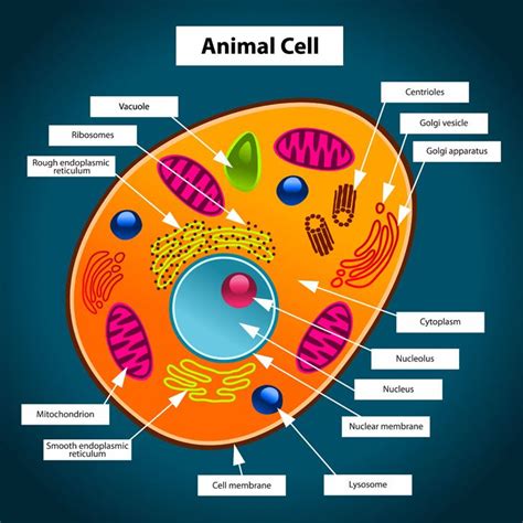
Table of Contents
A Deep Dive into Animal Cell Structure: A Visual Guide with Detailed Labels
Understanding the intricacies of animal cells is fundamental to grasping the complexities of biology. This comprehensive guide provides a detailed exploration of animal cell structure, accompanied by a labeled diagram for enhanced comprehension. We'll delve into the functions of each organelle, their interconnectedness, and the overall workings of this essential unit of life. This in-depth analysis will equip you with a strong foundation for further biological study.
The Animal Cell: A Microscopic Marvel
Animal cells, the building blocks of animal tissues and organs, are eukaryotic cells, meaning they possess a membrane-bound nucleus containing the genetic material (DNA). Unlike plant cells, they lack a rigid cell wall and chloroplasts. This absence allows for greater flexibility and a wider range of shapes and sizes. However, the absence of a cell wall doesn't mean animal cells are less complex; they are incredibly sophisticated entities with a variety of organelles working in concert to maintain cellular function.
Key Components of the Animal Cell: A Labeled Diagram Overview
Before diving into the detailed description of each component, let's first visualize the key structures of an animal cell. Imagine a well-organized factory, with each organelle representing a specialized department performing a specific task. (Note: A visual representation of a labeled animal cell diagram would be included here in a real blog post. Since I cannot create images, I will describe the key components and their locations within a hypothetical diagram).
The diagram would showcase the following components and their relative positions:
- Central Location: The nucleus, often the largest organelle, would be centrally located.
- Surrounding the Nucleus: The endoplasmic reticulum (ER), a network of interconnected membranes, would appear as a series of interconnected sacs and tubules extending from the nuclear envelope.
- Close Proximity to the ER: Ribosomes, small granular structures, would be shown both free-floating in the cytoplasm and attached to the rough ER.
- Near the Nucleus (and potentially throughout the cytoplasm): The Golgi apparatus (or Golgi body), a stack of flattened membrane-bound sacs, would be depicted.
- Scattered throughout the cytoplasm: Mitochondria, often depicted as bean-shaped organelles, would be distributed across the cytoplasm.
- Within the Cytoplasm: Lysosomes, small, membrane-bound organelles, would be scattered.
- Throughout the Cytoplasm (but perhaps less prominent visually): Peroxisomes would be shown as small, membrane-bound sacs.
- Enclosing the entire cell: The cell membrane (plasma membrane) would form the outer boundary.
- Inside the Nucleus: The nucleolus, a darkly stained region, would be visible within the nucleus.
- Within the Cytoplasm (potentially less easily visible without magnification): Centrosomes and centrioles would be depicted near the nucleus, particularly important during cell division.
- Throughout the Cytoplasm (often not easily visible without specific staining): Cytoskeleton filaments would be represented throughout the cell to show their supportive structure.
Detailed Description of Animal Cell Organelles
Now, let's explore each organelle in detail:
1. Nucleus: The Control Center
The nucleus, the cell's control center, houses the cell's genetic material, DNA, organized into chromosomes. It dictates cellular activities by regulating gene expression. The nuclear envelope, a double membrane perforated by nuclear pores, controls the passage of molecules between the nucleus and the cytoplasm. Within the nucleus, the nucleolus, a dense region, is responsible for ribosome synthesis.
2. Ribosomes: Protein Factories
Ribosomes are responsible for protein synthesis. These tiny organelles translate the genetic code from mRNA into proteins. They can be free-floating in the cytoplasm or bound to the endoplasmic reticulum. Free ribosomes produce proteins used within the cytoplasm, while ribosomes bound to the ER synthesize proteins for export or incorporation into membranes.
3. Endoplasmic Reticulum (ER): The Manufacturing and Transport Network
The endoplasmic reticulum (ER) is a vast network of interconnected membranes forming sacs and tubules. It's divided into two types:
- Rough ER: studded with ribosomes, it's involved in protein synthesis, folding, and modification.
- Smooth ER: lacks ribosomes and plays a role in lipid synthesis, detoxification, and calcium storage.
4. Golgi Apparatus: The Processing and Packaging Center
The Golgi apparatus (or Golgi body) receives proteins and lipids from the ER, further processes, modifies, sorts, and packages them into vesicles for transport to their final destinations within or outside the cell. Think of it as the cell's post office.
5. Mitochondria: The Powerhouses
Mitochondria are the cell's powerhouses, generating ATP (adenosine triphosphate), the cell's primary energy currency, through cellular respiration. They possess their own DNA and ribosomes, suggesting an endosymbiotic origin.
6. Lysosomes: The Waste Recyclers
Lysosomes are membrane-bound organelles containing digestive enzymes. They break down waste materials, cellular debris, and pathogens, maintaining cellular cleanliness and recycling cellular components.
7. Peroxisomes: Detoxification Specialists
Peroxisomes are involved in various metabolic processes, notably the breakdown of fatty acids and detoxification of harmful substances like hydrogen peroxide.
8. Cell Membrane (Plasma Membrane): The Gatekeeper
The cell membrane (or plasma membrane) is the outer boundary of the cell, regulating the passage of substances into and out of the cell. It's selectively permeable, meaning it controls which molecules can cross.
9. Centrosomes and Centrioles: Essential for Cell Division
Centrosomes, located near the nucleus, are microtubule-organizing centers. They contain centrioles, cylindrical structures crucial for cell division, playing a key role in organizing the mitotic spindle.
10. Cytoskeleton: The Cell's Internal Framework
The cytoskeleton is a network of protein filaments that provides structural support, maintains cell shape, and facilitates intracellular transport. It consists of microtubules, microfilaments, and intermediate filaments.
Interconnectedness of Organelles: A Symphony of Cellular Activities
The organelles within an animal cell don't function in isolation; they are intricately interconnected, working together in a coordinated fashion to maintain cellular homeostasis and perform various cellular functions. For example, proteins synthesized by ribosomes on the rough ER are transported to the Golgi apparatus for further processing before being delivered to their final destinations. Mitochondria provide the energy required for these processes. Lysosomes break down waste products, and the cytoskeleton provides structural support for the entire operation.
Conclusion: A Deeper Understanding of Cellular Life
Understanding the structure and function of animal cells is crucial for comprehending the complexities of life at the cellular level. This detailed guide, coupled with a visual representation of a labeled animal cell diagram, provides a solid foundation for further exploration into cell biology and related fields. Remember to consult additional resources and engage in further research to expand your knowledge and appreciation for the intricate world of animal cells. The more you delve into this subject, the more fascinating and intricate the cellular mechanisms become. This detailed overview serves as a springboard for continued learning and exploration in the dynamic realm of cell biology.
Latest Posts
Latest Posts
-
What Can 51 Be Divided By
Mar 28, 2025
-
What Occupies Space And Has Mass
Mar 28, 2025
-
Equation Of A Line Parallel To The Y Axis
Mar 28, 2025
-
Choose The Best Reagents To Complete The Reaction Shown Below
Mar 28, 2025
-
Do Generators Produce Ac Or Dc
Mar 28, 2025
Related Post
Thank you for visiting our website which covers about Picture Of A Animal Cell With Labels . We hope the information provided has been useful to you. Feel free to contact us if you have any questions or need further assistance. See you next time and don't miss to bookmark.
