Diagram Of An Animal Cell With Labels
Juapaving
Mar 25, 2025 · 7 min read
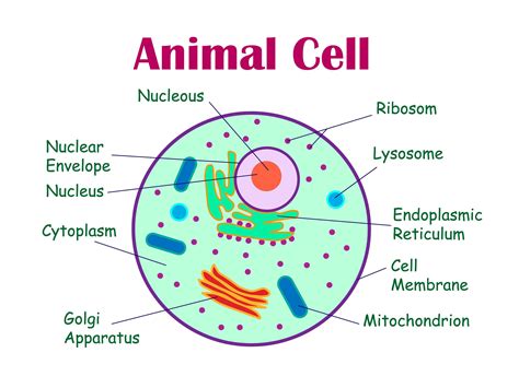
Table of Contents
A Deep Dive into the Animal Cell: A Diagram with Detailed Labels and Explanations
The animal cell, a fundamental unit of life, is a complex and fascinating structure brimming with activity. Understanding its intricate components is key to comprehending the processes of life itself. This comprehensive guide provides a detailed diagram of an animal cell, complete with labels for each organelle, and in-depth explanations of their functions. We'll explore the interconnectedness of these organelles and how they work together to maintain cellular homeostasis and support the overall function of the organism.
The Animal Cell: A Visual Representation
(Imagine a detailed diagram of an animal cell here. This would ideally be a high-quality image showing the nucleus, nucleolus, ribosomes, rough endoplasmic reticulum, smooth endoplasmic reticulum, Golgi apparatus, mitochondria, lysosomes, peroxisomes, centrosomes, cytoplasm, and cell membrane clearly labeled.)
While a physical diagram is impossible to include in this text format, I strongly encourage you to search for "animal cell diagram labeled" on Google Images to find a high-quality visual aid to complement this text. Referencing such a diagram will significantly enhance your understanding as you read through the descriptions below.
Key Components of the Animal Cell: A Detailed Breakdown
Let's delve into the specific organelles within the animal cell, exploring their structure and function in detail:
1. Cell Membrane (Plasma Membrane)
The cell membrane, or plasma membrane, is the outermost boundary of the animal cell. It's a selectively permeable barrier, meaning it controls what enters and exits the cell. This crucial function is performed by a phospholipid bilayer, a double layer of phospholipid molecules arranged with their hydrophilic (water-loving) heads facing outwards and their hydrophobic (water-fearing) tails facing inwards. Embedded within this bilayer are various proteins, which serve numerous functions, including transport of molecules, cell signaling, and cell adhesion. The membrane's fluidity allows for dynamic changes in its composition and function. The selective permeability of the cell membrane is essential for maintaining the internal environment of the cell, a process crucial for cell survival and function.
2. Cytoplasm
The cytoplasm is the jelly-like substance filling the cell between the cell membrane and the nucleus. It's composed mainly of water, salts, and various organic molecules. The cytoplasm is the site of many metabolic reactions, providing a medium for the organelles to interact and function. The cytoskeleton, a network of protein filaments, is also found within the cytoplasm, providing structural support and facilitating intracellular transport. This dynamic environment is crucial for numerous cellular processes, including protein synthesis and cellular respiration.
3. Nucleus
The nucleus, often referred to as the "control center" of the cell, houses the cell's genetic material, DNA. The DNA is organized into chromosomes, which carry the instructions for building and maintaining the cell. The nucleus is enclosed by a double membrane called the nuclear envelope, which is perforated by nuclear pores that regulate the passage of molecules between the nucleus and the cytoplasm. Inside the nucleus, the nucleolus is a prominent structure responsible for ribosome synthesis. The nucleus's role in regulating gene expression is vital for cellular differentiation, growth, and reproduction.
4. Ribosomes
Ribosomes are the protein synthesis factories of the cell. These tiny organelles are composed of RNA and protein and can be found free-floating in the cytoplasm or attached to the endoplasmic reticulum. Their primary function is to translate the genetic information encoded in mRNA (messenger RNA) into proteins, following the instructions provided by the nucleus. The efficiency and accuracy of ribosomes are essential for cellular growth, repair, and overall function.
5. Endoplasmic Reticulum (ER)
The endoplasmic reticulum (ER) is an extensive network of interconnected membranes extending throughout the cytoplasm. There are two types of ER:
-
Rough Endoplasmic Reticulum (RER): Studded with ribosomes, the RER plays a critical role in protein synthesis and modification. Proteins synthesized on the RER are often destined for secretion or integration into cellular membranes. The RER also helps in the folding and modification of these proteins, ensuring their proper function.
-
Smooth Endoplasmic Reticulum (SER): Lacking ribosomes, the SER is involved in lipid synthesis, carbohydrate metabolism, and detoxification. It plays a vital role in producing steroids, phospholipids, and other essential lipids. The SER also helps regulate calcium levels within the cell, crucial for numerous cellular processes.
6. Golgi Apparatus (Golgi Body)
The Golgi apparatus acts as the cell's processing and packaging center. It receives proteins and lipids synthesized in the ER, modifies them, and sorts them into vesicles for transport to their final destinations, either within the cell or for secretion outside the cell. This organelle plays a crucial role in the formation of lysosomes and other secretory vesicles. The Golgi's highly organized structure ensures efficient processing and targeted delivery of cellular products.
7. Mitochondria
Often referred to as the "powerhouses" of the cell, mitochondria are responsible for cellular respiration. These double-membraned organelles are the sites of ATP (adenosine triphosphate) production, the primary energy currency of the cell. The inner membrane of the mitochondria is folded into cristae, increasing the surface area for ATP synthesis. Mitochondria also play a role in cell signaling and apoptosis (programmed cell death). Their efficiency in energy production is vital for all cellular functions.
8. Lysosomes
Lysosomes are membrane-bound organelles containing hydrolytic enzymes that break down waste materials and cellular debris. They act as the cell's recycling centers, digesting unwanted substances and pathogens. Lysosomes maintain cellular homeostasis by removing damaged organelles and recycling their components. The controlled action of their enzymes is crucial for preventing cellular damage and maintaining cell health.
9. Peroxisomes
Peroxisomes are small, membrane-bound organelles that contain enzymes involved in various metabolic reactions. They play a crucial role in the breakdown of fatty acids and detoxification of harmful substances, including hydrogen peroxide. Peroxisomes protect the cell from oxidative damage by neutralizing reactive oxygen species. Their function in lipid metabolism and detoxification is essential for cellular health.
10. Centrosomes
Located near the nucleus, the centrosomes are microtubule-organizing centers, playing a key role in cell division. They contain a pair of centrioles, cylindrical structures that are involved in the formation of the mitotic spindle, which separates chromosomes during cell division. The centrosome's precise function in orchestrating cell division is crucial for the accurate transmission of genetic information during replication.
11. Vacuoles
While less prominent in animal cells than in plant cells, vacuoles are membrane-bound sacs that store various substances, including water, nutrients, and waste products. They contribute to maintaining cellular turgor pressure and can participate in cellular processes like endocytosis and exocytosis.
Interconnectedness of Organelles: A Cellular Symphony
The organelles within an animal cell don't operate in isolation; they are intricately interconnected and work together in a coordinated manner. For instance, the nucleus provides instructions for protein synthesis, which is carried out by ribosomes, often located on the RER. Proteins are then modified and packaged by the Golgi apparatus and transported to their final destinations. Mitochondria provide the energy required for these processes, while lysosomes break down cellular waste and damaged organelles. This coordinated effort highlights the remarkable efficiency and complexity of the animal cell.
Conclusion: The Marvel of the Animal Cell
The animal cell is a microcosm of life's complexity, a testament to the elegance and efficiency of biological design. Understanding its intricate structure and the functions of its various organelles is essential for comprehending a wide range of biological processes, from cellular respiration to protein synthesis to cell division. This detailed examination of the animal cell, accompanied by a visual diagram, provides a foundation for further exploration of the fascinating world of cell biology. Remember to consult a labeled diagram for a complete visual understanding. The more you delve into the intricacies of the animal cell, the more you’ll appreciate the wonder of life at its most fundamental level.
Latest Posts
Latest Posts
-
All Of The Multiples Of 7
Mar 26, 2025
-
What Is Not A Pure Substance
Mar 26, 2025
-
Lowest Common Denominator Of 7 And 9
Mar 26, 2025
-
Label The Cross Section Of A Leaf
Mar 26, 2025
-
Difference Between Plant Mitosis And Animal Mitosis
Mar 26, 2025
Related Post
Thank you for visiting our website which covers about Diagram Of An Animal Cell With Labels . We hope the information provided has been useful to you. Feel free to contact us if you have any questions or need further assistance. See you next time and don't miss to bookmark.
