Diagram Of An Animal Cell And A Plant Cell
Juapaving
Mar 20, 2025 · 5 min read
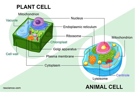
Table of Contents
Delving Deep: A Comparative Look at Animal and Plant Cell Diagrams
Understanding the fundamental building blocks of life—cells—is crucial for grasping the intricacies of biology. While all cells share certain characteristics, significant differences exist between plant and animal cells. These differences are reflected in their structures and ultimately, their functions. This comprehensive guide will delve into the detailed diagrams of both animal and plant cells, highlighting their key components and explaining their roles. We'll explore the similarities and differences, offering a thorough understanding of these vital cellular units.
Understanding the Basic Cell Structure: A Shared Foundation
Before diving into the specifics of plant and animal cells, let's establish a common ground. Both plant and animal cells are eukaryotic cells, meaning they possess a membrane-bound nucleus containing their genetic material (DNA). This nucleus acts as the control center, regulating cellular activities. Both cell types also share several essential organelles:
Shared Organelles:
-
Cell Membrane (Plasma Membrane): This selectively permeable membrane encloses the cell's contents, regulating the passage of substances in and out. Think of it as the cell's gatekeeper, controlling what enters and exits. Its structure, a phospholipid bilayer with embedded proteins, is vital for maintaining homeostasis within the cell.
-
Cytoplasm: The cytoplasm is the jelly-like substance filling the cell. It's not just an empty space; it's a dynamic environment where many cellular processes occur. Organelles are suspended within the cytoplasm, and it's the site of many metabolic reactions.
-
Ribosomes: These are the protein factories of the cell. They're responsible for synthesizing proteins based on the instructions encoded in the DNA. Ribosomes can be found free-floating in the cytoplasm or attached to the endoplasmic reticulum.
-
Endoplasmic Reticulum (ER): This extensive network of membranes plays a crucial role in protein and lipid synthesis. There are two types: rough ER (studded with ribosomes) and smooth ER (lacking ribosomes). The rough ER modifies and transports proteins, while the smooth ER synthesizes lipids and detoxifies certain substances.
-
Golgi Apparatus (Golgi Body): This organelle acts as the cell's processing and packaging center. It receives proteins and lipids from the ER, modifies them, and sorts them into vesicles for transport to their final destinations within or outside the cell. Think of it as the cell's postal service.
-
Mitochondria: These are often called the "powerhouses" of the cell because they're responsible for cellular respiration. This process generates ATP (adenosine triphosphate), the primary energy currency of the cell. Mitochondria have their own DNA, suggesting an endosymbiotic origin.
-
Lysosomes: These membrane-bound sacs contain digestive enzymes that break down waste materials, cellular debris, and even invading pathogens. They're crucial for maintaining cellular cleanliness and recycling cellular components.
-
Vacuoles: These are membrane-bound sacs used for storage. They can store water, nutrients, waste products, and other substances. The size and function of vacuoles differ significantly between plant and animal cells.
Diagram of an Animal Cell: A Detailed Exploration
An animal cell diagram reveals a complex interplay of organelles working together. While the shared organelles mentioned above are present, certain structures are unique or more prominent in animal cells.
(Insert a detailed and labelled diagram of an animal cell here. The diagram should clearly show the cell membrane, cytoplasm, nucleus, ribosomes, endoplasmic reticulum (rough and smooth), Golgi apparatus, mitochondria, lysosomes, centrioles, and possibly a few vacuoles. Use clear labels and different colors to distinguish the various organelles.)
Animal Cell-Specific Structures:
-
Centrioles: These cylindrical structures play a crucial role in cell division, organizing microtubules during mitosis and meiosis. They are typically found in pairs near the nucleus.
-
Small Vacuoles: Animal cells have numerous small vacuoles compared to the large central vacuole found in plant cells. These vacuoles perform various functions, including storage and waste removal.
Diagram of a Plant Cell: Unique Features and Adaptations
Plant cells exhibit many features not found in animal cells, reflecting their specialized functions. The most striking difference lies in the presence of a rigid cell wall and a large central vacuole.
(Insert a detailed and labelled diagram of a plant cell here. The diagram should clearly show the cell membrane, cell wall, cytoplasm, nucleus, ribosomes, endoplasmic reticulum (rough and smooth), Golgi apparatus, mitochondria, chloroplasts, large central vacuole, and plasmodesmata. Use clear labels and different colors to distinguish the various organelles.)
Plant Cell-Specific Structures:
-
Cell Wall: This rigid outer layer provides structural support and protection to the plant cell. It's primarily composed of cellulose, a complex carbohydrate. The cell wall maintains the cell's shape and prevents excessive water uptake.
-
Chloroplasts: These are the sites of photosynthesis, the process by which plants convert light energy into chemical energy in the form of glucose. Chloroplasts contain chlorophyll, the green pigment that absorbs light energy. They also have their own DNA, suggesting an endosymbiotic origin, similar to mitochondria.
-
Large Central Vacuole: This prominent vacuole occupies a significant portion of the plant cell's volume. It plays a vital role in maintaining turgor pressure, storing water, nutrients, and waste products. The large central vacuole contributes significantly to the cell's size and shape.
-
Plasmodesmata: These are channels that connect adjacent plant cells, allowing for communication and transport of substances between cells. They're essential for coordinating activities across the plant tissue.
Comparing Animal and Plant Cells: A Side-by-Side Analysis
| Feature | Animal Cell | Plant Cell |
|---|---|---|
| Cell Wall | Absent | Present (cellulose) |
| Chloroplasts | Absent | Present (chlorophyll) |
| Vacuoles | Small, numerous | Large, central vacuole |
| Centrioles | Present | Usually absent |
| Shape | Variable, often irregular | Typically rectangular or polygonal |
| Size | Generally smaller | Generally larger |
| Photosynthesis | Absent | Present |
| Plasmodesmata | Absent | Present (connecting adjacent cells) |
Conclusion: A Deeper Appreciation for Cellular Diversity
This detailed exploration of animal and plant cell diagrams reveals the complexity and diversity within the cellular world. While both cell types share fundamental characteristics, their unique features reflect their distinct roles and adaptations. Understanding these differences is fundamental to appreciating the remarkable diversity of life on Earth. By grasping the structure and function of each organelle, we gain a deeper understanding of how these microscopic units contribute to the overall functioning of organisms. Further exploration into cellular biology will continue to reveal more intricate details and refine our understanding of these fundamental building blocks of life.
Latest Posts
Latest Posts
-
Multiplication Chart From 1 To 20
Mar 21, 2025
-
What Are 3 Fractions Equivalent To 3 8
Mar 21, 2025
-
Highest Common Factor Of 40 And 25
Mar 21, 2025
-
What Is 42 C In Fahrenheit
Mar 21, 2025
-
Intermediate Value Theorem Vs Mean Value Theorem
Mar 21, 2025
Related Post
Thank you for visiting our website which covers about Diagram Of An Animal Cell And A Plant Cell . We hope the information provided has been useful to you. Feel free to contact us if you have any questions or need further assistance. See you next time and don't miss to bookmark.
