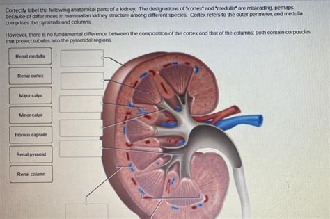
Correctly Label The Following Anatomical Parts Of A Kidney.
Juapaving
Apr 05, 2025 · 7 min read

Table of Contents
Correctly Labeling the Anatomical Parts of a Kidney: A Comprehensive Guide
The kidney, a vital organ in the urinary system, plays a crucial role in maintaining overall health by filtering blood and removing waste products. Understanding its intricate anatomy is fundamental to comprehending its complex functions. This comprehensive guide will delve into the detailed anatomical structures of the kidney, providing a clear and concise explanation for correct labeling. We'll explore both macroscopic and microscopic aspects, equipping you with a thorough understanding of this remarkable organ.
Macroscopic Anatomy of the Kidney: External Structures
Let's begin by examining the external features visible to the naked eye. A healthy kidney resembles a reddish-brown bean, approximately the size of a fist. Several key external structures are essential for proper labeling:
1. Renal Capsule: The Protective Outer Layer
The renal capsule is a tough, fibrous membrane that directly encloses the kidney. This protective layer acts as a barrier against infection and physical trauma, safeguarding the delicate internal structures. Its smooth, glistening surface contributes to the overall appearance of the kidney. Correct labeling requires identifying this outermost covering.
2. Renal Fascia: Anchoring the Kidney
Surrounding the renal capsule is the renal fascia, a thin layer of connective tissue. This fascia helps to anchor the kidney in place, preventing excessive movement within the abdominal cavity. While not directly part of the kidney itself, its presence is crucial for proper positioning and overall stability.
3. Perirenal Fat: Cushioning and Protection
Enveloping the renal fascia is the perirenal fat, a substantial layer of adipose tissue. This fatty cushion provides significant protection to the kidney, absorbing shocks and preventing damage from external forces. Its thickness can vary depending on the individual's overall body fat percentage.
4. Hilum: The Gateway to the Kidney
The hilum is a concave medial border where several important structures enter and exit the kidney. This indentation serves as a gateway for the renal artery, renal vein, and ureter, facilitating the intricate process of blood filtration and urine excretion. Precise labeling of the hilum is crucial in understanding the kidney's vascular and urinary connections.
Macroscopic Anatomy of the Kidney: Internal Structures
Moving beyond the external features, let's explore the complex internal structures of the kidney. These intricate components work together in a coordinated fashion to perform the vital functions of blood filtration and waste removal.
1. Renal Cortex: The Outer Region
The renal cortex is the outer region of the kidney, characterized by a granular appearance. This area contains the functional units of the kidney, the nephrons, responsible for the initial stages of urine formation. The cortex is vital for filtration and reabsorption, contributing significantly to the kidney's overall function. Accurate labeling requires distinguishing it from the deeper medulla.
2. Renal Medulla: The Inner Region
Deep to the cortex lies the renal medulla, a darker, striated region. The medulla is composed of cone-shaped structures called renal pyramids. These pyramids contain the loops of Henle and collecting ducts, which play crucial roles in concentrating urine. The striated appearance is due to the parallel arrangement of these structures. Correctly identifying the medulla and its pyramids is essential for understanding urine concentration mechanisms.
3. Renal Columns: Extensions of the Cortex
Extending from the cortex into the medulla are the renal columns, also known as the columns of Bertin. These cortical extensions separate the renal pyramids, providing structural support and maintaining the overall architecture of the kidney. Their presence is important for maintaining the integrity of the medullary structures.
4. Renal Papillae: Urine Collection Points
The apex of each renal pyramid is the renal papilla, which projects into a minor calyx. These papillae act as the points of urine collection, marking the transition from the medulla to the collecting system. The urine flows from the papillae into the minor calyces. Accurate labeling necessitates identifying the papillae as the culmination of urine processing within the pyramids.
5. Minor Calyces: The Initial Collecting Units
Several minor calyces collect urine from the renal papillae. These cup-like structures are the initial components of the renal pelvis, initiating the process of urine transportation towards the ureter. Labeling the minor calyces requires understanding their role as the first stage of urine collection within the kidney.
6. Major Calyces: Combining Urine Flow
Multiple minor calyces merge to form larger major calyces. These structures further collect and channel urine, funneling it towards the next stage in the urinary tract. Correct labeling clarifies the progressive collection and consolidation of urine.
7. Renal Pelvis: Urine Collection Reservoir
The renal pelvis is a funnel-shaped structure formed by the convergence of major calyces. It serves as a reservoir for urine before its passage into the ureter. The renal pelvis is a crucial landmark in understanding the flow of urine from the kidney's interior to the urinary bladder. Accurate labeling involves recognizing its central position within the kidney's drainage system.
8. Ureter: Transporting Urine to the Bladder
The ureter is a muscular tube that emerges from the renal pelvis and transports urine to the urinary bladder. Its peristaltic contractions propel urine along its length, ensuring efficient movement of waste products out of the kidney. Labeling the ureter signifies the final stage of urine transport within the kidney's anatomical context.
Microscopic Anatomy of the Kidney: The Nephron
Delving into the microscopic realm, the functional unit of the kidney is the nephron. Millions of nephrons reside within each kidney, tirelessly working to filter blood and produce urine. The nephron's intricate structure is essential for understanding the detailed mechanisms of urine formation. Let's explore its key components:
1. Renal Corpuscle: The Filtration Unit
The renal corpuscle is the initial filtering component of the nephron. It consists of two main structures:
a) Glomerulus: The Capillary Network
The glomerulus is a network of specialized capillaries where blood is initially filtered. The high pressure within these capillaries facilitates the passage of water and small molecules into Bowman's capsule. Precise labeling highlights its crucial role in the initial stages of filtration.
b) Bowman's Capsule: Enclosing the Glomerulus
Bowman's capsule is a double-walled cup-like structure that surrounds the glomerulus. It receives the filtrate from the glomerulus, initiating the journey of this fluid towards the formation of urine. Accurate labeling emphasizes its role as the initial recipient of the glomerular filtrate.
2. Renal Tubule: Processing the Filtrate
The renal tubule is a long, convoluted tube that processes the filtrate received from Bowman's capsule. Several distinct segments contribute to the complex processes of reabsorption and secretion:
a) Proximal Convoluted Tubule (PCT): Reabsorption of Nutrients
The proximal convoluted tubule (PCT) is the initial segment of the renal tubule. It's responsible for the reabsorption of essential nutrients, such as glucose, amino acids, and water, returning them to the bloodstream. Accurate labeling highlights its importance in selective reabsorption.
b) Loop of Henle: Concentrating Urine
The loop of Henle is a U-shaped structure that extends into the renal medulla. It plays a critical role in concentrating urine by establishing an osmotic gradient, allowing for the reabsorption of water. Correct labeling emphasizes its significance in urine concentration.
c) Distal Convoluted Tubule (DCT): Fine-tuning the Filtrate
The distal convoluted tubule (DCT) is the final segment of the renal tubule. It fine-tunes the composition of the filtrate, regulating electrolyte balance and pH. Accurate labeling points out its role in the precise adjustment of urine composition.
3. Collecting Duct: Final Urine Formation
Multiple distal convoluted tubules converge into a collecting duct. These ducts collect urine from multiple nephrons and transport it to the renal papillae. The collecting ducts play a role in regulating water reabsorption, contributing to the final concentration of urine. Precise labeling emphasizes its role in the final stages of urine formation.
Conclusion: Mastering Kidney Anatomy
Correctly labeling the anatomical parts of a kidney necessitates a thorough understanding of both macroscopic and microscopic structures. From the protective renal capsule to the intricate nephron, each component plays a crucial role in the kidney's vital functions. By mastering the details outlined in this guide, you'll gain a deeper appreciation for this remarkable organ's complexity and its indispensable contribution to overall health. This knowledge forms a solid foundation for further exploration into the physiology and pathophysiology of the urinary system. Remember to utilize various learning resources, including diagrams, models, and interactive simulations, to enhance your comprehension and solidify your ability to accurately label each anatomical part.
Latest Posts
Latest Posts
-
Which Of The Following Is Not A Property Of Bases
Apr 06, 2025
-
What Is Difference Between Gas And Vapour
Apr 06, 2025
-
Words That Begin And End With R
Apr 06, 2025
-
What Is The Tangent Of 30 Degrees
Apr 06, 2025
-
What Distinguishes An Element From A Compound
Apr 06, 2025
Related Post
Thank you for visiting our website which covers about Correctly Label The Following Anatomical Parts Of A Kidney. . We hope the information provided has been useful to you. Feel free to contact us if you have any questions or need further assistance. See you next time and don't miss to bookmark.
