Why The Wall Of The Left Ventricle Is Thicker
Juapaving
Mar 14, 2025 · 5 min read
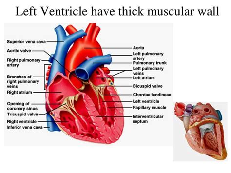
Table of Contents
Why the Left Ventricular Wall is Thicker: A Deep Dive into Cardiac Anatomy and Physiology
The human heart, a tireless engine driving life's processes, is a marvel of biological engineering. Within its four chambers, a complex interplay of pressure, volume, and muscular force ensures the continuous circulation of oxygenated blood throughout the body. One striking anatomical feature is the significantly greater thickness of the left ventricular wall compared to the right. This difference isn't arbitrary; it's a direct consequence of the vastly different workloads these chambers endure. This article will delve into the intricate reasons behind this crucial anatomical disparity, exploring the physiological demands placed on each ventricle and the resulting adaptations in myocardial structure.
The Functional Differences Between the Left and Right Ventricles
To understand why the left ventricular wall is thicker, we must first appreciate the fundamental differences in the functions of the left and right ventricles. The heart functions as a dual pump, with each side handling a distinct circulatory pathway:
-
Right Ventricle: This chamber receives deoxygenated blood from the body via the superior and inferior vena cava and pumps it to the lungs for oxygenation. The pulmonary circulation, the pathway to and from the lungs, is a relatively low-pressure system. The resistance to blood flow is minimal.
-
Left Ventricle: This chamber receives oxygenated blood from the lungs via the pulmonary veins and pumps it to the entire body through the aorta. This systemic circulation involves pumping blood against significantly higher resistance. The aorta branches into a vast network of arteries supplying every organ and tissue, requiring substantially more pressure.
The Impact of Systemic Vascular Resistance
The key difference lies in the systemic vascular resistance (SVR). SVR represents the resistance the blood encounters as it flows through the systemic circulation. This resistance is substantially higher than the pulmonary vascular resistance, primarily due to the significantly smaller diameter and longer length of systemic arteries compared to pulmonary arteries. To overcome this higher resistance and propel blood throughout the body, the left ventricle must generate considerably more pressure.
Pressure-Volume Relationship and Myocardial Hypertrophy
This higher pressure requirement translates directly into a need for greater muscular force. The left ventricle achieves this through myocardial hypertrophy, a process of increased muscle mass. The thicker wall allows for stronger contractions, enabling the left ventricle to generate the high pressures necessary for effective systemic blood circulation. This is a prime example of form following function – the physiological demands of the systemic circulation directly shape the anatomical structure of the left ventricle.
The Role of the Frank-Starling Law
The Frank-Starling law of the heart further illuminates the importance of left ventricular thickness. This law states that the stroke volume of the heart is directly proportional to the initial length of the cardiac muscle fibers (preload). A larger left ventricle, with its thicker wall and greater chamber volume, can accommodate a larger end-diastolic volume (the volume of blood in the ventricle at the end of diastole). This increased preload allows for a more powerful contraction and a larger stroke volume, enhancing the efficiency of the left ventricle in overcoming systemic vascular resistance.
Microscopic Anatomy and Cellular Adaptations
The increased thickness of the left ventricular wall is not simply an increase in the number of muscle cells; there are also significant changes at the cellular level. Myocardial cells in the left ventricle are larger and contain a higher density of contractile proteins (actin and myosin) compared to those in the right ventricle. This increased protein density allows for a more forceful contraction with each heartbeat.
Furthermore, the left ventricular myocardium exhibits a more organized arrangement of cardiomyocytes, further enhancing contractile efficiency. The specific arrangement of collagen fibers within the ventricular wall also plays a role in providing structural support against the high pressures generated during systole (ventricular contraction).
Clinical Implications of Left Ventricular Hypertrophy
While left ventricular hypertrophy is a natural adaptation to the demands of systemic circulation, excessive or pathological hypertrophy can have significant clinical implications. Conditions like hypertension (high blood pressure), aortic stenosis (narrowing of the aortic valve), and hypertrophic cardiomyopathy (a genetic disorder) can lead to significant left ventricular thickening, ultimately impairing cardiac function. This can result in:
- Heart failure: The thickened ventricle may become less efficient at pumping blood, leading to fluid accumulation in the lungs (pulmonary edema) and other parts of the body.
- Arrhythmias: The altered electrical properties of the hypertrophied myocardium can increase the risk of abnormal heart rhythms, potentially leading to life-threatening conditions.
- Sudden cardiac death: In severe cases, particularly those associated with hypertrophic cardiomyopathy, excessive left ventricular hypertrophy can increase the risk of sudden cardiac death.
Comparative Anatomy and Evolutionary Considerations
The difference in ventricular wall thickness is not unique to humans; it's observed across various vertebrate species. The degree of hypertrophy in the left ventricle is often correlated with the animal's metabolic rate and the demands of its systemic circulation. For instance, highly active animals often exhibit more pronounced left ventricular hypertrophy compared to less active species. This reflects the evolutionary adaptation of the heart to the unique physiological demands of different species.
Diagnostic Assessment of Left Ventricular Function
Various diagnostic tools are used to assess left ventricular function and detect any abnormalities. These include:
- Echocardiography: A non-invasive ultrasound technique provides detailed images of the heart's structure and function, allowing for accurate measurement of left ventricular wall thickness and assessment of contractile function.
- Cardiac MRI: Offers high-resolution images of the heart, providing more detailed information about the structure and function of the left ventricle.
- Cardiac Catheterization: An invasive procedure allowing for direct measurement of left ventricular pressures and assessment of coronary artery health.
Conclusion
The significantly thicker wall of the left ventricle is a testament to the body's remarkable ability to adapt to physiological demands. This anatomical difference is not a mere quirk; it's a crucial adaptation that enables the heart to effectively pump blood against the high resistance of the systemic circulation. Understanding the underlying mechanisms driving this hypertrophy is essential for comprehending normal cardiac physiology and diagnosing and managing various cardiovascular diseases. Further research continues to unravel the complexities of cardiac remodeling and the intricate interplay between genetics, environment, and the development of heart disease. The continued exploration of left ventricular function remains a critical area of investigation in cardiovascular medicine, offering hope for improved diagnosis, treatment, and ultimately, prevention of cardiac-related morbidity and mortality.
Latest Posts
Latest Posts
-
How Many Black Cards In A Deck Of 52
Mar 14, 2025
-
Are The Diagonals In A Parallelogram Perpendicular
Mar 14, 2025
-
What Is 2 10 As A Percent
Mar 14, 2025
-
What Is The Horizontal Row In The Periodic Table
Mar 14, 2025
-
What Mirror Provides The Widest Field Of View
Mar 14, 2025
Related Post
Thank you for visiting our website which covers about Why The Wall Of The Left Ventricle Is Thicker . We hope the information provided has been useful to you. Feel free to contact us if you have any questions or need further assistance. See you next time and don't miss to bookmark.
