Which Phase Do Chromosomes First Become Visible
Juapaving
Apr 01, 2025 · 5 min read
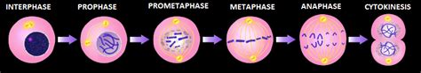
Table of Contents
Which Phase Do Chromosomes First Become Visible? A Deep Dive into Cell Division
Understanding when chromosomes first become visible is crucial to grasping the fundamental processes of cell division – mitosis and meiosis. These processes are vital for growth, repair, and reproduction in all living organisms. This article will explore the intricacies of chromosome visibility, delving into the specific stages of cell division where these structures first become discernible under a microscope, and clarifying common misconceptions.
The Cell Cycle: A Preparatory Stage
Before we pinpoint the exact phase, it's essential to understand the broader context: the cell cycle. The cell cycle is a series of events that lead to cell growth and division. It consists of two major phases: interphase and the mitotic (or meiotic) phase. Interphase, often mistakenly considered a "resting" phase, is actually a period of intense activity where the cell grows, replicates its DNA, and prepares for division.
Interphase: A Period of Growth and DNA Replication
Interphase itself is subdivided into three stages:
-
G1 (Gap 1) phase: The cell grows in size, produces RNA and synthesizes proteins. Chromosomes are present, but they are not yet condensed and therefore are not individually visible under a light microscope. They appear as a diffuse mass of chromatin.
-
S (Synthesis) phase: DNA replication occurs. Each chromosome duplicates itself, creating two identical sister chromatids joined at the centromere. Again, the chromosomes remain uncondensed and indistinguishable under a light microscope. The duplicated genetic material remains dispersed as chromatin.
-
G2 (Gap 2) phase: The cell continues to grow and prepare for mitosis or meiosis. Further protein synthesis takes place, ensuring the cell has all the necessary components for chromosome segregation. Chromosomes are still uncondensed and invisible as individual structures under a standard light microscope.
The Mitotic Phase: Where Chromosomes Become Visible
The mitotic (M) phase is where the replicated genetic material is accurately segregated into two daughter cells. This phase encompasses several distinct stages:
Prophase: The Beginning of Chromosome Condensation
It is during prophase that chromosomes first become visible under a light microscope. The lengthy, thread-like chromatin fibers begin to condense, coiling and compacting into shorter, thicker structures. This condensation process makes individual chromosomes distinguishable. Each chromosome now consists of two identical sister chromatids joined at the centromere. While visible, they are not yet fully condensed to their most compact form.
Key Prophase Events Leading to Chromosome Visibility:
-
Chromatin Condensation: The complex process involving histone proteins and other factors facilitates the tight packaging of DNA. This compaction reduces the length of the DNA molecule dramatically, making the chromosomes observable under the microscope.
-
Nuclear Envelope Breakdown: The nuclear envelope, which encloses the nucleus, starts to break down. This allows the condensed chromosomes to move freely into the cytoplasm, preparing for their segregation.
-
Spindle Fiber Formation: Microtubules begin to assemble, forming the mitotic spindle. The spindle fibers will later attach to the chromosomes, playing a vital role in their segregation.
Metaphase: Maximum Chromosome Condensation and Alignment
While chromosomes are visible in prophase, their condensation continues into metaphase. Metaphase represents the stage where chromosomes reach their maximum degree of condensation and are most easily observed under a light microscope. They align themselves along the metaphase plate, an imaginary plane equidistant from the two poles of the cell. This alignment is crucial for ensuring accurate chromosome segregation.
Metaphase: The Peak of Visibility
The tightly coiled and condensed state of chromosomes in metaphase allows for clear visualization under a microscope. This is the stage where karyotyping, a technique used to study chromosome number and structure, is typically performed. The distinct morphology of each chromosome – its size, shape, and banding pattern – is readily apparent.
Anaphase and Telophase: Decondensation Begins
After metaphase, the sister chromatids separate and move toward opposite poles of the cell during anaphase. Though still visible, their degree of condensation begins to slightly decrease. In telophase, this decondensation continues, and the chromosomes become less compact, eventually reverting to a dispersed chromatin state within the newly formed nuclei.
Meiosis: A Similar, Yet Distinct Process
Meiosis, the process of cell division that produces gametes (sperm and egg cells), also involves chromosome condensation and visibility. However, there are some key differences compared to mitosis:
Meiosis I: Reductional Division
-
Prophase I: This is the longest and most complex phase of meiosis. It is during prophase I that chromosomes first become visible, similar to mitosis. However, a unique event occurs: homologous chromosomes pair up (synapsis) and exchange genetic material through crossing over. This recombination shuffles genetic information, increasing genetic diversity.
-
Metaphase I: Homologous chromosome pairs align at the metaphase plate.
-
Anaphase I: Homologous chromosomes separate and move toward opposite poles. Sister chromatids remain attached.
Meiosis II: Equational Division
Meiosis II is very similar to mitosis. Chromosomes are already condensed and visible from the beginning of this phase. Sister chromatids separate during anaphase II, resulting in four haploid daughter cells.
Common Misconceptions and Clarifications
Several misconceptions surround the visibility of chromosomes:
-
Interphase is a “resting” phase: This is incorrect. Interphase is a period of intense activity, including DNA replication and cell growth. Chromosomes are present but not visible as distinct entities.
-
Chromosomes are always visible: Chromosomes are only visible under a light microscope during specific phases of cell division when they are condensed. During interphase, they exist as a dispersed chromatin network.
-
All microscopes show chromosomes clearly: The resolution of the microscope is crucial. A standard light microscope is sufficient to see condensed chromosomes, especially during metaphase.
Conclusion: Prophase Marks the Onset of Chromosome Visibility
In summary, chromosomes first become visible under a light microscope during prophase of both mitosis and meiosis. This is due to the process of chromatin condensation, which compacts the DNA into shorter, thicker structures. While visible in prophase, chromosomes reach their maximum condensation and are most clearly observable during metaphase. Understanding the cell cycle and the stages of cell division is essential for comprehending the fundamental processes of life and the mechanics of heredity. The detailed examination of chromosome behavior across these stages reveals the intricate choreography of genetic material manipulation underlying growth, repair, and the continuation of life.
Latest Posts
Latest Posts
-
Volume Of A Cylinder In Cubic Feet
Apr 02, 2025
-
69 Is The Square Root Of
Apr 02, 2025
-
Is The Number 13 Prime Or Composite
Apr 02, 2025
-
How Many Miles Is 2 5 Km
Apr 02, 2025
-
How Long Is 13 Cm In Inches
Apr 02, 2025
Related Post
Thank you for visiting our website which covers about Which Phase Do Chromosomes First Become Visible . We hope the information provided has been useful to you. Feel free to contact us if you have any questions or need further assistance. See you next time and don't miss to bookmark.
