What Phase Of Mitosis Is Pictured
Juapaving
Mar 23, 2025 · 6 min read
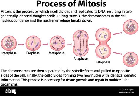
Table of Contents
What Phase of Mitosis is Pictured? A Comprehensive Guide to Identifying Mitosis Stages
Identifying the specific phase of mitosis from a microscopic image requires careful observation and understanding of the key characteristics of each stage. Mitosis, the process of cell division resulting in two identical daughter cells, unfolds in a series of distinct phases: prophase, prometaphase, metaphase, anaphase, and telophase. This guide will help you confidently determine the phase depicted in a given microscopic image, focusing on the critical visual cues that differentiate each stage. We'll also explore the significance of accurate identification in various scientific contexts.
Understanding the Stages of Mitosis
Before delving into identification, let's review the defining features of each mitotic phase:
1. Prophase: The Beginning of Chromosome Condensation
Prophase is characterized by the initial condensation of chromatin into visible chromosomes. Each chromosome consists of two identical sister chromatids joined at the centromere. While the nuclear envelope remains intact during early prophase, it begins to fragment as prophase progresses. The mitotic spindle, composed of microtubules, starts to form outside the nucleus, originating from centrosomes which have begun migrating to opposite poles of the cell. Nucleoli, the sites of ribosome synthesis, become less distinct or disappear entirely. Looking for these key features—condensed chromosomes, fragmented nuclear envelope, and forming mitotic spindle—is crucial for identifying prophase.
2. Prometaphase: Nuclear Envelope Breakdown and Chromosome Attachment
Prometaphase marks the complete breakdown of the nuclear envelope. This allows the spindle microtubules to interact directly with the chromosomes. Kinetochores, protein structures located at the centromeres, become attached to the microtubules. This attachment is crucial for the subsequent alignment of chromosomes at the metaphase plate. The chromosomes undergo further condensation, becoming even more compact and distinct. To distinguish prometaphase, look for the absence of a nuclear envelope, chromosomes interacting with the spindle microtubules, and kinetochores actively attaching.
3. Metaphase: Chromosomes Align at the Equator
Metaphase is characterized by the precise alignment of chromosomes at the metaphase plate, an imaginary plane equidistant from the two spindle poles. Each chromosome's kinetochores are attached to microtubules from both poles, ensuring equal segregation of sister chromatids during the subsequent anaphase. This alignment is a critical checkpoint in mitosis; the cell doesn't proceed until all chromosomes are correctly positioned. The key visual characteristic of metaphase is the perfect alignment of chromosomes along the metaphase plate. The chromosomes are highly condensed and clearly visible.
4. Anaphase: Sister Chromatids Separate
Anaphase marks the dramatic separation of sister chromatids. The centromeres split, and each chromatid, now considered an independent chromosome, is pulled towards opposite poles of the cell by the shortening of the kinetochore microtubules. Simultaneously, the non-kinetochore microtubules lengthen, pushing the poles further apart, elongating the cell. The hallmark of anaphase is the visible separation of sister chromatids moving towards opposite poles. The cell elongates noticeably.
5. Telophase: Formation of Two Nuclei
Telophase is essentially the reverse of prophase. The chromosomes arrive at the poles and begin to decondense, becoming less visible. The nuclear envelope reforms around each set of chromosomes, creating two separate nuclei. The mitotic spindle disassembles. Cytokinesis, the division of the cytoplasm, typically overlaps with telophase. The key identifiers of telophase are the decondensing chromosomes, reforming nuclear envelopes, and the appearance of two distinct nuclei.
Identifying the Phase: A Step-by-Step Approach
Analyzing a microscopic image to determine the mitotic phase requires a systematic approach. Here's a step-by-step guide:
-
Assess Chromosome Condensation: Are the chromosomes highly condensed and easily distinguishable (metaphase), moderately condensed (prophase, prometaphase), or decondensed and diffuse (telophase)?
-
Examine the Nuclear Envelope: Is the nuclear envelope intact (prophase, early prophase), fragmented (prometaphase), or completely absent (metaphase, anaphase)?
-
Observe the Mitotic Spindle: Is the mitotic spindle forming (prophase), interacting with chromosomes (prometaphase, metaphase, anaphase), or disassembling (telophase)?
-
Check Chromosome Alignment: Are the chromosomes aligned at the metaphase plate (metaphase)? Are sister chromatids separating (anaphase)?
-
Note the Presence of Two Nuclei: Are two distinct nuclei forming (telophase)?
By systematically evaluating these features, you can accurately identify the phase of mitosis depicted in the microscopic image.
The Importance of Accurate Identification
Accurate identification of mitotic phases is crucial in various biological and medical contexts:
-
Cancer Research: Analyzing the mitotic index (the percentage of cells undergoing mitosis) and the fidelity of mitosis are essential in cancer research, as uncontrolled or aberrant cell division is a hallmark of cancer. Understanding the specific mitotic phase helps assess the effectiveness of anticancer drugs.
-
Developmental Biology: The precise regulation of mitosis is vital for proper embryonic development. Studying mitotic phases helps uncover mechanisms underlying developmental processes and identify potential causes of developmental abnormalities.
-
Genetic Research: Observing mitosis allows scientists to study chromosome segregation and the transmission of genetic material during cell division. Identifying specific phases helps pinpoint errors in chromosome segregation that can lead to genetic disorders.
-
Plant Biology: Mitosis in plants has some unique characteristics. Accurately identifying phases is crucial for understanding the growth and development of plants and for optimizing agricultural practices.
Common Pitfalls and How to Avoid Them
Several factors can make identifying the mitotic phase challenging:
-
Image Quality: Poor image resolution or artifacts can obscure crucial details, making accurate identification difficult. Ensure you're using high-quality microscopy images.
-
Cell Type and Species: The appearance of mitosis can vary slightly between different cell types and species. Be aware of these variations and consult appropriate references.
-
Overlap of Phases: The transition between phases is gradual, and sometimes, cells might display characteristics of two adjacent phases. Careful consideration of all features is crucial in such cases.
Advanced Techniques for Mitosis Analysis
While basic light microscopy is sufficient for identifying mitotic phases in many cases, advanced techniques offer higher resolution and detailed insights:
-
Fluorescence Microscopy: Using fluorescently labeled proteins allows visualization of specific structures involved in mitosis, such as microtubules and kinetochores, providing a more detailed picture of the process.
-
Time-lapse Microscopy: Live-cell imaging allows observation of mitosis in real-time, revealing dynamic changes occurring during the different phases.
-
3D Microscopy: Advanced microscopy techniques enable the construction of 3D models of cells, offering a more comprehensive view of mitotic structures and their spatial organization.
Conclusion
Accurately identifying the phase of mitosis depicted in a microscopic image is a fundamental skill for anyone working in biology or related fields. By carefully observing the key characteristics of each phase – chromosome condensation, nuclear envelope integrity, spindle formation, chromosome alignment, and sister chromatid separation – and applying a systematic approach, you can confidently determine the mitotic stage. Understanding the significance of accurate identification is essential for advancing our knowledge of cell biology, genetics, and various related areas. Mastering this skill will empower you to contribute to various scientific endeavors and medical advancements.
Latest Posts
Latest Posts
-
Whats The Lcm Of 8 And 10
Mar 25, 2025
-
What Is 11 Cm In Inches
Mar 25, 2025
-
Least Common Multiple Of 40 And 15
Mar 25, 2025
-
What Base Is Found In Rna But Not Dna
Mar 25, 2025
-
What Is The Least Common Multiple Of 12 And 16
Mar 25, 2025
Related Post
Thank you for visiting our website which covers about What Phase Of Mitosis Is Pictured . We hope the information provided has been useful to you. Feel free to contact us if you have any questions or need further assistance. See you next time and don't miss to bookmark.
