What Color Is The Animal Cell
Juapaving
Mar 09, 2025 · 6 min read
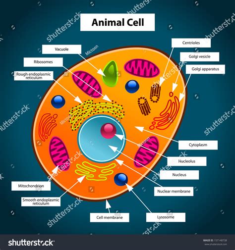
Table of Contents
What Color Is the Animal Cell? Exploring the Nuances of Cellular Coloration
The question, "What color is an animal cell?" might seem deceptively simple. However, the answer reveals a fascinating exploration into the complexities of cell biology, microscopy, and the very nature of color perception. The straightforward answer is: animal cells have no inherent color. Their appearance under a microscope is largely dictated by the staining techniques used and the presence of specific pigments or organelles. Let's delve deeper into the factors that contribute to the perceived color of animal cells.
The Invisible World: Unstained Animal Cells
In their natural, unstained state, animal cells are largely translucent or colorless. This is because the cytoplasm, the jelly-like substance filling the cell, lacks significant pigmentation. Individual cellular components, such as the nucleus, mitochondria, and endoplasmic reticulum, are also transparent. Viewing these cells under a light microscope without staining would reveal only faint outlines and possibly some internal structures depending on the cell type and the microscope's resolution. This lack of inherent color makes staining techniques essential for visualizing cell structures and functions.
The Power of Staining: Unveiling Cellular Structures
Staining techniques are crucial for revealing the intricate details of animal cells. These methods involve using dyes that bind to specific cellular components, making them visible under a microscope. Different dyes bind to different structures, allowing researchers to distinguish between various organelles and cellular processes. The color we perceive is directly related to the dye used and its interaction with the cell.
Common Staining Techniques and Their Color Effects:
-
Hematoxylin and Eosin (H&E): This is perhaps the most ubiquitous staining method in histology. Hematoxylin stains nucleic acids (DNA and RNA) a deep purple or blue, highlighting the nucleus and other structures rich in nucleic acids. Eosin, on the other hand, stains cytoplasmic proteins pink or red. Thus, using H&E staining, we observe a predominantly pink and purple image, with the nucleus standing out prominently in shades of blue-purple.
-
Wright-Giemsa Stain: Frequently used in hematology (the study of blood), this stain differentiates blood cells based on their morphology and chemical composition. It produces a range of colors, with nuclei staining dark purple or blue-black, cytoplasm staining various shades of pink, purple, and blue, and granules within cells showing different colors depending on their chemical nature.
-
Sudan Black B: This stain is specifically used to identify lipids (fats) within cells, staining them a black or dark blue color. This is vital for observing lipid droplets and other lipid-rich structures.
-
Periodic Acid-Schiff (PAS) Stain: This stain detects carbohydrates and glycoproteins, coloring these structures a bright magenta or pink. This is particularly useful in studying the glycocalyx, the carbohydrate-rich layer surrounding many cells.
-
Immunofluorescence Staining: This advanced technique employs fluorescently labeled antibodies that bind to specific proteins or antigens within the cell. The resulting colors depend entirely on the fluorescent dye used, ranging from bright green, red, blue, yellow, and other spectral colors, allowing researchers to simultaneously visualize multiple cellular components.
Beyond Staining: Factors Affecting Perceived Cell Color
While staining techniques significantly influence the perceived color, other factors can also contribute:
-
Cell Type: Different cell types naturally contain varying amounts of pigments or metabolites that can subtly affect their appearance. For instance, some cells might contain pigments like melanin (responsible for skin and hair color), which could impart a brown or black hue even without staining.
-
Specimen Preparation: The way cells are prepared for microscopy can also impact their color. Factors such as fixation techniques and embedding media can subtly alter the way the cells interact with stains, leading to variations in color intensity and distribution.
-
Microscope Settings: The microscope's settings, such as illumination intensity and filter selection, play a significant role in how the stained cells are perceived. Adjustments in these settings can alter the brightness and contrast of the observed colors.
-
Optical Properties: The refractive index of the cellular components also influences light transmission and scattering, affecting the overall appearance. This is often less about color and more about clarity and contrast but contributes to the overall visual experience.
The Importance of Accurate Interpretation
It's crucial to remember that the color observed in stained animal cells is an artifact of the staining process rather than a reflection of the cells' natural coloration. The choice of staining technique depends entirely on the research objective – each method is designed to highlight specific cellular components or processes. Therefore, interpreting the color of cells in microscopy requires a thorough understanding of the staining procedure and the biological context.
Applications in Medical Diagnostics and Research
The coloration of animal cells, as revealed through staining, plays a critical role in various applications:
-
Disease Diagnosis: Pathologists routinely use staining techniques to diagnose diseases. Abnormal cell morphology and staining patterns can indicate cancerous or infectious processes. For example, specific staining patterns can help distinguish different types of leukemia.
-
Drug Development and Testing: Cell-based assays often employ staining methods to evaluate the effectiveness of new drugs. The ability of a drug to affect cell viability, morphology, or specific cellular processes can be assessed by observing changes in cell staining patterns.
-
Basic Biological Research: Understanding cellular structures and functions relies heavily on accurate visualization using staining techniques. Researchers use various staining methods to study cell division, apoptosis (programmed cell death), and other fundamental biological processes.
Advanced Microscopy Techniques: Moving Beyond Traditional Staining
While staining remains a vital tool, advancements in microscopy have provided alternative methods for visualizing animal cells without relying solely on dyes:
-
Phase-Contrast Microscopy: This technique enhances the contrast between different cellular components without staining, allowing for observation of unstained cells. While not producing "color" in the traditional sense, it reveals differences in refractive index, producing a grayscale image with enhanced contrast.
-
Differential Interference Contrast (DIC) Microscopy: Similar to phase-contrast microscopy, DIC enhances contrast by exploiting differences in refractive index. This results in a three-dimensional appearance of the cell, providing detailed structural information without the need for staining.
-
Confocal Microscopy: This sophisticated technique allows for the creation of high-resolution images of cells by eliminating out-of-focus light. While often used with fluorescent staining, it can also be employed with other contrast-enhancing techniques.
-
Electron Microscopy: This technique provides incredibly high-resolution images of cells, revealing ultrastructural details that are invisible under light microscopy. Electron microscopy doesn't involve staining in the same way as light microscopy but uses heavy metal staining techniques for contrast.
Conclusion: The Dynamic World of Cellular Coloration
The question of an animal cell's color is far more nuanced than it initially appears. While animal cells are naturally colorless and translucent, the use of staining techniques and various microscopy methods brings their intricate structures into view. The perceived color is entirely dependent on the chosen method and the specific cellular components being visualized. Understanding the color imparted by various stains is crucial for accurate interpretation and has profound implications in medical diagnostics, drug development, and fundamental biological research. The dynamic interplay of staining, microscopy techniques, and cellular structures continues to shape our understanding of the unseen world within living organisms.
Latest Posts
Latest Posts
-
How Do You Calculate The Perimeter Of A Triangle
Mar 10, 2025
-
Which Of The Following Statements Is Incorrect
Mar 10, 2025
-
In What Organelle Does Cellular Respiration Occur
Mar 10, 2025
-
What Is The Squar Root Of 49
Mar 10, 2025
-
How Many Valence Electrons Does K Have
Mar 10, 2025
Related Post
Thank you for visiting our website which covers about What Color Is The Animal Cell . We hope the information provided has been useful to you. Feel free to contact us if you have any questions or need further assistance. See you next time and don't miss to bookmark.
