These Neurons Transmit Impulses From Cns To Effectors
Juapaving
Mar 30, 2025 · 7 min read
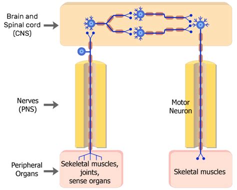
Table of Contents
Motor Neurons: The Messengers of Movement from CNS to Effectors
Motor neurons are the crucial link between the central nervous system (CNS), which encompasses the brain and spinal cord, and the body's effectors. Effectors are essentially the working parts of the body—muscles and glands—that respond to nervous impulses. These neurons transmit impulses from the CNS, initiating actions like muscle contractions, glandular secretions, and other bodily responses. Understanding their function is vital for comprehending movement, reflexes, and overall bodily control. This in-depth exploration will delve into the intricate world of motor neurons, examining their structure, types, neurotransmission process, and the critical role they play in maintaining homeostasis and coordinating complex behaviours.
The Structure of Motor Neurons
Motor neurons are classified as efferent neurons, meaning they carry signals away from the CNS. Unlike sensory neurons, which transmit signals toward the CNS, motor neurons initiate actions. Their structure is specialized for this efferent function. A typical motor neuron comprises several key components:
1. Soma (Cell Body):
The soma is the neuron's metabolic center, containing the nucleus and other organelles essential for cell maintenance and function. It integrates incoming signals from other neurons and determines whether to transmit a signal further. The soma's location varies depending on the motor neuron type; in some, it resides within the CNS, while in others, it lies outside the CNS within ganglia.
2. Dendrites:
These branched extensions of the soma receive incoming signals from other neurons, primarily interneurons within the CNS. The numerous dendritic branches significantly increase the surface area for receiving these signals. The integration of these incoming signals determines whether the neuron will fire an action potential.
3. Axon:
The axon is a long, slender projection extending from the soma. It's responsible for transmitting the nerve impulse (action potential) away from the soma to the effector organ. The axon's length can vary significantly, ranging from short connections within the spinal cord to extremely long axons that reach muscles in the extremities. The axon is often covered by a myelin sheath, which greatly increases the speed of nerve impulse transmission.
4. Axon Terminals (Synaptic Terminals):
At the axon's end are numerous branches called axon terminals, each ending in a synaptic terminal (or bouton). These terminals form synapses with the effector cells (muscle fibers or gland cells). The synapse is the junction where the motor neuron communicates with the effector cell, releasing neurotransmitters to initiate a response.
Types of Motor Neurons
Motor neurons aren't a monolithic group; they exhibit significant diversity based on their function and location within the nervous system. Two primary classifications exist:
1. Alpha Motor Neurons (α-motor neurons):
These are the most abundant type of motor neuron and directly innervate skeletal muscle fibers. Each α-motor neuron forms neuromuscular junctions with several muscle fibers, collectively called a motor unit. The strength of a muscle contraction is determined by the number of motor units recruited. These neurons are responsible for voluntary movements. Their large diameter and myelination contribute to the speed of their signal transmission, ensuring rapid muscle responses. The excitation of an α-motor neuron results in the contraction of the muscle fibers it innervates.
2. Gamma Motor Neurons (γ-motor neurons):
These motor neurons innervate intrafusal muscle fibers within muscle spindles. Muscle spindles are sensory receptors embedded within muscles that monitor muscle length and rate of change in length. γ-motor neurons adjust the sensitivity of muscle spindles, maintaining optimal muscle tone and proprioception (awareness of body position and movement). Their function is crucial for maintaining muscle responsiveness and coordinating precise movements. Unlike α-motor neurons, γ-motor neurons play a crucial role in feedback mechanisms regulating muscle activity.
Neurotransmission at the Neuromuscular Junction
The process by which motor neurons transmit impulses to effectors is called neurotransmission. This process unfolds at specialized junctions called synapses, specifically neuromuscular junctions in the case of muscle innervation. The steps are as follows:
-
Action Potential Arrival: An action potential travels down the axon of the motor neuron, reaching the axon terminal.
-
Calcium Influx: The arrival of the action potential triggers voltage-gated calcium channels in the axon terminal to open. Calcium ions (Ca²⁺) rush into the axon terminal.
-
Vesicle Fusion: The influx of Ca²⁺ initiates a cascade of events leading to the fusion of synaptic vesicles with the presynaptic membrane. These vesicles contain the neurotransmitter acetylcholine (ACh).
-
Acetylcholine Release: ACh is released into the synaptic cleft, the narrow gap between the axon terminal and the muscle fiber's motor end plate.
-
Acetylcholine Binding: ACh diffuses across the synaptic cleft and binds to nicotinic acetylcholine receptors on the motor end plate.
-
Depolarization: ACh binding opens ligand-gated ion channels, allowing sodium ions (Na⁺) to enter the muscle fiber. This influx of Na⁺ depolarizes the muscle fiber membrane, initiating an action potential.
-
Muscle Contraction: The action potential spreads along the muscle fiber's sarcolemma, triggering the release of calcium ions from the sarcoplasmic reticulum. This leads to the interaction of actin and myosin filaments, resulting in muscle contraction.
-
Acetylcholine Breakdown: The action of ACh is terminated by the enzyme acetylcholinesterase (AChE), which breaks down ACh into choline and acetate. This prevents continuous muscle contraction. Choline is then transported back into the axon terminal to be resynthesized into ACh.
Motor Neuron Disorders and Diseases
Several neurological disorders affect motor neuron function, leading to debilitating symptoms. Understanding these conditions is crucial for developing effective treatments. Some of the prominent examples include:
1. Amyotrophic Lateral Sclerosis (ALS):
ALS, also known as Lou Gehrig's disease, is a progressive neurodegenerative disease affecting motor neurons in the brain and spinal cord. This leads to muscle weakness, atrophy, and eventually paralysis. The cause of ALS is unknown, although genetic factors and environmental toxins are considered potential contributors.
2. Spinal Muscular Atrophy (SMA):
SMA is a group of inherited disorders characterized by the degeneration of motor neurons in the spinal cord. This results in progressive muscle weakness and atrophy, affecting voluntary movement. The severity of SMA varies depending on the specific gene mutation.
3. Poliomyelitis (Polio):
Polio is a viral infection that can damage motor neurons, causing paralysis. While largely eradicated globally due to vaccination efforts, polio remains a threat in some regions.
4. Multiple Sclerosis (MS):
MS is an autoimmune disease that affects the myelin sheath surrounding nerve fibers, including motor neurons. Damage to the myelin sheath disrupts nerve impulse transmission, leading to various neurological symptoms, including muscle weakness and fatigue.
The Importance of Motor Neurons in Complex Movements
The seemingly effortless execution of complex movements relies on the intricate coordination of numerous motor neurons working in concert. The brain, through its complex neural networks, orchestrates this coordination. Consider playing a piano: the precise finger movements, the coordinated activation of different muscles, and the timing of each note press all stem from the precise activation patterns of motor neurons directed by the brain's motor cortex and cerebellum.
Conclusion
Motor neurons are essential components of the nervous system, acting as the final common pathway for the execution of voluntary and involuntary movements and glandular secretions. Their structural specialization, diverse types, and crucial role in neurotransmission at the neuromuscular junction underscore their importance in maintaining homeostasis and facilitating a wide range of bodily functions. Understanding their function and the consequences of their dysfunction is vital for diagnosing, treating, and researching a wide range of neurological disorders. The complexity of their role in coordinated movement highlights the intricate sophistication of the nervous system and its remarkable ability to orchestrate a seamless interaction between the brain and the body's effectors. Further research into the molecular mechanisms underlying motor neuron function and the pathogenesis of motor neuron diseases holds the key to developing new therapeutic strategies and improving the lives of those affected by these debilitating conditions.
Latest Posts
Latest Posts
-
What Is The Multiple Of 15
Apr 01, 2025
-
The Proper Electron Dot Symbol For Aluminum Is
Apr 01, 2025
-
Biuret Test Shows The Presence Of
Apr 01, 2025
-
What Is The Distance Between Rarefactions Called
Apr 01, 2025
-
13 Gallons Is How Many Liters
Apr 01, 2025
Related Post
Thank you for visiting our website which covers about These Neurons Transmit Impulses From Cns To Effectors . We hope the information provided has been useful to you. Feel free to contact us if you have any questions or need further assistance. See you next time and don't miss to bookmark.
