The Protein Coat Of A Virus Is Called
Juapaving
Mar 31, 2025 · 6 min read
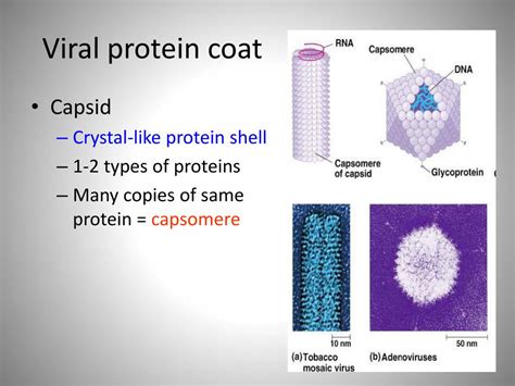
Table of Contents
The Protein Coat of a Virus: A Deep Dive into the Capsid and Its Importance
The protein coat of a virus is called a capsid. This isn't just a simple shell; it's a complex and meticulously structured entity crucial for viral survival and infectivity. Understanding the capsid's structure, function, and variations is fundamental to comprehending virology and developing effective antiviral strategies. This article will delve deep into the world of viral capsids, exploring their composition, assembly, diversity, and significance in viral pathogenesis.
What is a Capsid?
A capsid is the protein shell that encloses the genetic material (either DNA or RNA) of a virus. Think of it as the virus's protective armor and delivery system. It's composed of numerous protein subunits called capsomeres. These capsomeres self-assemble into highly organized structures, forming a protective container that shields the viral genome from degradation by enzymes and other environmental factors. The capsid's structure is not merely protective; it plays a critical role in the virus's ability to attach to and infect host cells.
Capsomere: The Building Blocks of the Capsid
Capsomere structure varies greatly depending on the virus. They are often made of multiple polypeptide chains, and their precise arrangement dictates the overall architecture of the capsid. The interaction between capsomeres is primarily driven by non-covalent bonds, such as hydrogen bonds, hydrophobic interactions, and electrostatic forces. This allows for relatively easy assembly and disassembly of the capsid, which is important for the viral life cycle.
Types of Capsid Structures
Viral capsids are broadly classified into two main categories based on their symmetry:
1. Helical Capsids
Helical capsids are characterized by a rod-like or filamentous structure. The capsomeres arrange themselves in a spiral pattern around the viral nucleic acid, creating a tube-like structure. This type of capsid is commonly found in viruses that infect plants and animals, such as tobacco mosaic virus (TMV) and influenza viruses (although influenza viruses also have an envelope). The length of a helical capsid is often proportional to the length of the viral genome.
2. Icosahedral Capsids
Icosahedral capsids are more complex and exhibit a high degree of symmetry. An icosahedron is a 20-faced geometric solid, and the capsomeres are arranged in a precise manner to create this symmetrical structure. This arrangement is highly efficient, allowing for the enclosure of a relatively large volume of genetic material with a minimal number of capsomeres. Many animal and plant viruses, including adenoviruses and human papillomaviruses (HPVs), possess icosahedral capsids. The arrangement and number of capsomeres can vary, leading to different sizes and appearances of icosahedral capsids.
Variations and Combinations
While helical and icosahedral structures are the most common, some viruses display more complex capsid architectures. These include:
- Complex Capsids: Certain viruses, like bacteriophages (viruses that infect bacteria), possess complex capsids that combine elements of both helical and icosahedral symmetries. These often have a head region (icosahedral) and a tail region (helical) involved in attaching to and injecting genetic material into the host cell. This complex structure reflects the intricate mechanisms these viruses use to infect their specific bacterial hosts.
The Role of the Capsid in Viral Infection
The capsid is not merely a passive container; it actively participates in every stage of viral infection:
1. Attachment to Host Cells
The capsid plays a crucial role in the initial attachment of the virus to the host cell. Specific proteins on the capsid surface, known as viral attachment proteins, bind to complementary receptors on the surface of the host cell. This interaction is highly specific, determining the host range of the virus—which types of cells it can infect. The precise binding between the viral attachment proteins and host cell receptors is a key determinant of viral tropism. Mutations in either the viral attachment proteins or the host cell receptors can dramatically alter the infectivity of the virus.
2. Entry into Host Cells
Following attachment, the virus needs to enter the host cell. The mechanism of entry varies greatly depending on the virus and its capsid structure. Some viruses enter via receptor-mediated endocytosis, where the host cell engulfs the virus in a vesicle. Others use direct fusion with the host cell membrane, releasing their genetic material directly into the cytoplasm. The capsid often undergoes conformational changes to facilitate entry, sometimes triggered by changes in pH or other environmental cues. Understanding these entry mechanisms is crucial for developing antiviral drugs that target the entry process.
3. Uncoating and Genome Release
Once inside the host cell, the virus must release its genome. This process is known as uncoating. This typically involves the disassembly of the capsid, either partially or completely. The specific mechanism of uncoating depends on the virus, often involving the interaction of viral proteins with host cell components. The release of the viral genome is essential for the virus to initiate its replication cycle within the host cell.
Capsid and Viral Evolution
The capsid is a primary target for the host immune system. Consequently, viral capsid proteins are under constant selective pressure to evolve, evading the immune response. This leads to antigenic variation, where mutations in the capsid proteins change their surface characteristics, making them less recognizable to antibodies. Influenza viruses are a prime example, exhibiting significant antigenic drift and shift in their hemagglutinin (HA) and neuraminidase (NA) surface glycoproteins, which are components of their envelope (a lipid membrane surrounding the capsid). This continuous evolution poses a major challenge for vaccine development.
Capsid Proteins and Antiviral Drug Targets
Given the crucial role of the capsid in the viral life cycle, its proteins represent attractive targets for antiviral drug development. These drugs can either directly inhibit capsid assembly or interfere with capsid-host cell interactions. Several antiviral drugs currently in use target viral capsid proteins, highlighting the importance of understanding capsid structure and function for therapeutic development.
Studying the Capsid: Techniques and Approaches
Researchers employ a variety of techniques to study viral capsids:
- X-ray crystallography: This technique allows for the determination of the three-dimensional structure of capsid proteins at high resolution. It provides invaluable insights into the arrangement of capsomeres and the interactions between them.
- Cryo-electron microscopy (cryo-EM): Cryo-EM is another powerful technique that provides high-resolution images of viral capsids, particularly useful for larger and more complex structures.
- Mass spectrometry: This technique is used to identify and characterize the protein components of the capsid.
- Biophysical techniques: Techniques such as dynamic light scattering and analytical ultracentrifugation are used to study the size, shape, and stability of capsids.
- Genetic techniques: Manipulating viral genes encoding capsid proteins allows for the study of the effects of mutations on capsid assembly, stability, and function. This provides valuable information about structure-function relationships.
Conclusion: The Capsid's Central Role in Virology
The capsid, the protein coat of a virus, is far more than a simple container. It's a sophisticated molecular machine that orchestrates every step of viral infection, from attachment to host cells to the release of the viral genome. Its remarkable structural diversity reflects the myriad strategies viruses have evolved to infect their hosts. Understanding the structure, assembly, function, and evolution of viral capsids remains a central focus of virology research, paving the way for the development of novel antiviral therapies and vaccines. Continued research in this area is essential to combat the ever-evolving threat of viral diseases. The intricacies of the capsid underscore the complexity and ingenuity of the viral world and highlight the importance of continuing to unlock its secrets.
Latest Posts
Latest Posts
-
What Are The Greatest Common Factors Of 75
Apr 02, 2025
-
Do Parallelograms Have 4 Equal Sides
Apr 02, 2025
-
What Is Group Of Baboons Called
Apr 02, 2025
-
Exterior Angle Of A Regular Octagon
Apr 02, 2025
-
What Are The Common Multiples Of 24
Apr 02, 2025
Related Post
Thank you for visiting our website which covers about The Protein Coat Of A Virus Is Called . We hope the information provided has been useful to you. Feel free to contact us if you have any questions or need further assistance. See you next time and don't miss to bookmark.
