The Pectoral Girdle Consists Of The
Juapaving
Apr 03, 2025 · 6 min read

Table of Contents
The Pectoral Girdle: A Comprehensive Overview of its Structure, Function, and Clinical Significance
The pectoral girdle, also known as the shoulder girdle, is a complex anatomical structure that plays a vital role in upper limb movement and overall body posture. Unlike the pelvic girdle, which is firmly attached to the axial skeleton, the pectoral girdle exhibits remarkable mobility, allowing for a wide range of arm movements. Understanding its intricate components, their interrelationships, and their clinical implications is crucial for healthcare professionals and anyone interested in human anatomy and biomechanics.
Components of the Pectoral Girdle: A Detailed Look
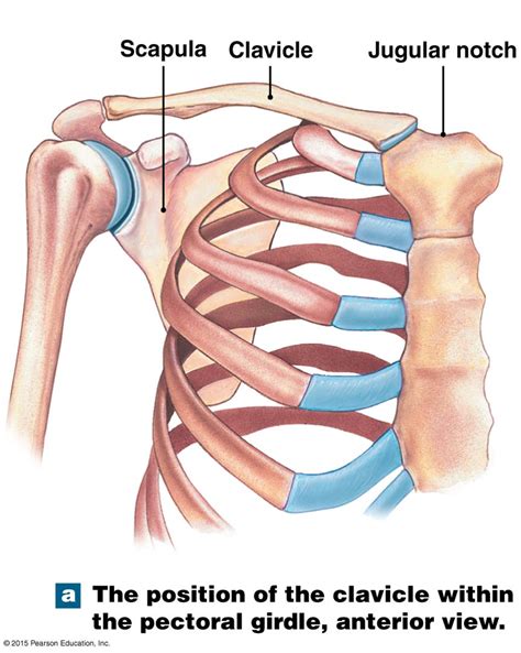
The pectoral girdle comprises two primary bones: the clavicle (collarbone) and the scapula (shoulder blade). These bones, along with their associated ligaments and muscles, work in concert to provide both stability and mobility to the shoulder joint.
The Clavicle: The Keystone of the Shoulder
The clavicle, a long S-shaped bone, is the only direct bony connection between the axial skeleton (the skull, vertebral column, and rib cage) and the upper limb. Its medial end articulates with the sternum at the sternoclavicular joint (SC joint), while its lateral end articulates with the acromion process of the scapula at the acromioclavicular joint (AC joint).
Key features of the clavicle:
- Medial (sternal) end: Thick and somewhat triangular, articulating with the manubrium of the sternum.
- Lateral (acromial) end: Flattened and broader, articulating with the acromion.
- Conoid tubercle: A small prominence on the inferior surface, serving as an attachment point for ligaments.
- Trapezoid line: A roughened area adjacent to the conoid tubercle, also providing ligament attachment.
The clavicle's unique shape and position are crucial for its function. It acts as a strut, transferring forces from the upper limb to the axial skeleton, protecting underlying neurovascular structures. Its mobility allows for a wide range of shoulder movements while providing stability.
The Scapula: The Versatile Shoulder Blade
The scapula is a large, flat, triangular bone located on the posterior aspect of the thorax. It doesn't directly articulate with the rib cage, but rather glides across its surface, providing exceptional mobility. This remarkable mobility is achieved through the interplay of several muscles that attach to the scapula.
Key features of the scapula:
- Spine: A prominent ridge running across the posterior surface, ending laterally in the acromion.
- Acromion: The lateral extension of the spine, articulating with the clavicle.
- Coracoid process: A hook-like projection on the anterior surface, serving as an attachment site for muscles.
- Glenoid cavity: A shallow, pear-shaped socket that articulates with the head of the humerus (upper arm bone), forming the glenohumeral joint.
- Suprascapular notch: A notch on the superior border, which transmits the suprascapular nerve.
- Subscapular fossa: A broad, concave surface on the anterior aspect, providing attachment for the subscapularis muscle.
- Infraspinous fossa: A concave surface below the spine, providing attachment for the infraspinatus muscle.
- Supraspinous fossa: A concave surface above the spine, providing attachment for the supraspinatus muscle.
The scapula’s versatility allows for a wide range of arm movements, including abduction, adduction, elevation, depression, upward and downward rotation. This adaptability is crucial for activities ranging from reaching for objects to throwing a ball.
Articulations of the Pectoral Girdle: The Joints that Enable Movement
The pectoral girdle's remarkable mobility is facilitated by several key joints:
-
Sternoclavicular (SC) Joint: This synovial joint, connecting the medial end of the clavicle to the manubrium of the sternum, allows for a combination of gliding and rotational movements. It is crucial for shoulder stability and range of motion.
-
Acromioclavicular (AC) Joint: This synovial joint, connecting the lateral end of the clavicle to the acromion process of the scapula, permits gliding and rotation. It plays a critical role in scapular movements.
-
Scapulothoracic Joint (Functional Joint): While not a true anatomical joint, the scapulothoracic articulation describes the gliding movement of the scapula across the posterior thoracic wall. This movement is facilitated by the muscles that attach to the scapula.
These joints, working in coordination with the surrounding muscles and ligaments, enable the complex and nuanced movements of the shoulder complex.
Muscles of the Pectoral Girdle: The Movers and Shapers
The pectoral girdle’s mobility depends heavily on the numerous muscles that attach to the clavicle and scapula. These muscles can be broadly categorized into those that primarily act on the scapula (scapulohumeral muscles) and those that connect the scapula to other parts of the body (intrinsic muscles of the shoulder). Understanding the action of each muscle is crucial for comprehending the overall mechanics of the shoulder.
Key muscles acting on the scapula:
- Trapezius: Elevates, retracts, and rotates the scapula.
- Levator scapulae: Elevates the scapula.
- Rhomboid major and minor: Retract and rotate the scapula.
- Serratus anterior: Protracts and rotates the scapula.
- Pectoralis minor: Depresses, protracts, and rotates the scapula.
These muscles, acting in concert, provide intricate control over scapular position and movement, facilitating a wide range of arm actions.
Clinical Significance of the Pectoral Girdle: Common Injuries and Conditions
The pectoral girdle's high degree of mobility makes it susceptible to a variety of injuries and conditions. Understanding these issues is crucial for diagnosis and treatment.
-
Clavicle Fractures: Commonly caused by falls or direct trauma, clavicle fractures are more frequent in younger individuals. Treatment may involve immobilization with a sling or surgical repair.
-
Acromioclavicular (AC) Joint Separation: This injury involves damage to the ligaments stabilizing the AC joint, often resulting from a fall onto the shoulder. Treatment ranges from conservative measures (sling and rest) to surgical reconstruction.
-
Rotator Cuff Injuries: The rotator cuff muscles (supraspinatus, infraspinatus, teres minor, and subscapularis) stabilize the shoulder joint and enable its movements. Tears in these muscles are common and can range from mild to severe, often requiring surgery.
-
Shoulder Dislocation: The glenohumeral joint, with its shallow socket, is prone to dislocation, typically anteriorly. Dislocations necessitate prompt reduction (realignment) and rehabilitation.
-
Subacromial Bursitis: Inflammation of the subacromial bursa, a fluid-filled sac located beneath the acromion, can cause pain and limited range of motion. Treatment may involve rest, anti-inflammatory medications, or corticosteroid injections.
-
Frozen Shoulder (adhesive capsulitis): This condition involves stiffness and pain in the shoulder joint, limiting range of motion. The cause isn't fully understood, but treatment focuses on restoring mobility through physical therapy.
-
Thoracic Outlet Syndrome: Compression of neurovascular structures in the thoracic outlet (space between the clavicle and first rib) can lead to pain, numbness, and weakness in the arm and hand.
-
Scapular Winging: This condition, characterized by the medial border of the scapula protruding from the back, is often caused by damage to the long thoracic nerve, leading to weakness in the serratus anterior muscle.
Understanding the anatomy and biomechanics of the pectoral girdle is essential for diagnosing and effectively managing these conditions.
Conclusion: The Pectoral Girdle – A Foundation of Upper Limb Function
The pectoral girdle, with its intricate network of bones, joints, and muscles, is a remarkable anatomical structure that enables the remarkable mobility and dexterity of the human upper limb. Its complex interplay of stability and mobility is crucial for a wide range of activities, from fine motor skills to strenuous physical tasks. The vulnerability of the shoulder to injury underscores the importance of understanding its anatomy, biomechanics, and clinical significance. This knowledge is vital for healthcare professionals and anyone interested in understanding the human body's intricate design and functionality. Further research continues to reveal deeper insights into the complexities of this pivotal region of the body, enhancing our understanding of injury mechanisms, treatment strategies, and preventive measures. Understanding the pectoral girdle's structure and function is key to promoting healthy movement and preventing injury.
Latest Posts
Latest Posts
-
As Temperature Increases The Rate Of Diffusion
Apr 03, 2025
-
Is 87 Prime Or Composite Number
Apr 03, 2025
-
Least Common Multiple 15 And 25
Apr 03, 2025
-
How Many Electrons Can Occupy The 3d Subshell
Apr 03, 2025
-
Difference Between And Enzyme And A Hormone
Apr 03, 2025
Related Post
Thank you for visiting our website which covers about The Pectoral Girdle Consists Of The . We hope the information provided has been useful to you. Feel free to contact us if you have any questions or need further assistance. See you next time and don't miss to bookmark.
