The Occipital Condyles Articulate With The
Juapaving
Apr 05, 2025 · 7 min read

Table of Contents
The Occipital Condyles: Articulation, Function, and Clinical Significance
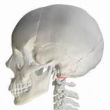
The occipital condyles are two oval-shaped bony projections located on the inferior surface of the occipital bone, at the base of the skull. Their primary function is to articulate with the superior articular facets of the first cervical vertebra, the atlas (C1), forming the atlanto-occipital joint. This crucial articulation allows for the nodding movement of the head – flexion and extension – and plays a vital role in maintaining head stability and facilitating a wide range of head and neck movements. Understanding the intricacies of this joint, including its articulation, function, and potential clinical implications, is crucial for healthcare professionals and anyone interested in human anatomy and biomechanics.
Anatomy of the Atlanto-Occipital Joint
The atlanto-occipital joint is a classic example of a diarthrodial or synovial joint. This means it’s characterized by the presence of a joint capsule, a synovial membrane that secretes lubricating synovial fluid, and articular cartilage covering the articulating surfaces. This sophisticated design allows for smooth, low-friction movement.
The Occipital Condyles: A Detailed Look
The occipital condyles themselves are slightly convex, mirroring the concave superior articular facets of the atlas. This reciprocal relationship contributes to the joint's stability. They are positioned laterally and slightly anteriorly on the occipital bone, contributing to the natural forward tilt of the head. The size and shape of the occipital condyles can vary slightly between individuals, which may influence the range of motion and susceptibility to injury.
The Atlas (C1): The Superior Articular Facets
The superior articular facets of the atlas are shaped to perfectly complement the occipital condyles. Their concave shape allows for a secure fit and facilitates a wide range of motion. These facets are oriented superiorly, medially, and slightly anteriorly, allowing for flexion and extension of the head. The arrangement of ligaments further enhances joint stability and restricts excessive movement.
Supporting Ligaments
Several crucial ligaments contribute to the stability and integrity of the atlanto-occipital joint:
-
Anterior Atlanto-Occipital Membrane: This strong, fibrous membrane runs from the anterior arch of the atlas to the anterior margin of the foramen magnum. It acts as a significant restraint against posterior displacement of the atlas relative to the occipital bone.
-
Posterior Atlanto-Occipital Membrane: A thinner membrane than its anterior counterpart, this ligament spans from the posterior arch of the atlas to the posterior margin of the foramen magnum. It offers a degree of posterior stability, although its contribution is less significant than the anterior membrane.
-
Lateral Atlanto-Occipital Ligaments: These robust ligaments connect the lateral masses of the atlas to the jugular processes of the occipital bone. They provide lateral stability and restrict lateral displacement of the atlas.
These ligaments work synergistically to prevent excessive movement and maintain the integrity of the atlanto-occipital joint. Damage or laxity in these ligaments can compromise joint stability and increase the risk of instability.
Function of the Atlanto-Occipital Joint
The primary function of the atlanto-occipital joint is to provide for the nodding movement of the head – flexion (tilting the head forward) and extension (tilting the head backward). This allows us to perform actions such as looking up, looking down, and acknowledging a greeting. While the joint's primary role is flexion and extension, it also allows for a limited amount of lateral flexion (side-to-side tilting) and rotation, although these movements are far more restricted compared to flexion-extension.
The sophisticated design of the atlanto-occipital joint, involving the precisely shaped articular surfaces, supporting ligaments, and synovial fluid, allows for a smooth, controlled range of motion. This precision is critical for maintaining head posture, gaze stability, and overall balance. The muscles that act upon this joint further refine these movements and contribute to the head and neck's overall control.
Muscles Involved in Atlanto-Occipital Joint Movement
Several muscles contribute to the movement and stability of the atlanto-occipital joint, including:
-
Rectus Capitis Anterior: This small muscle aids in flexion of the head.
-
Rectus Capitis Lateralis: Assists in lateral flexion of the head.
-
Rectus Capitis Posterior Major: Contributes to extension and slight rotation of the head.
-
Rectus Capitis Posterior Minor: Assists in extension and stabilization of the head.
-
Obliquus Capitis Superior: Contributes to extension and rotation of the head.
-
Obliquus Capitis Inferior: Assists in rotation of the head. This muscle is primarily involved in atlanto-axial joint movement but also indirectly influences the atlanto-occipital joint.
The coordinated action of these muscles allows for precise and controlled head movements. Weakness or dysfunction in these muscles can lead to impaired head control, neck pain, and postural abnormalities.
Clinical Significance and Potential Problems
The atlanto-occipital joint, while crucial for normal head movement, is also susceptible to injury and dysfunction. Understanding the potential problems associated with this joint is important for accurate diagnosis and appropriate management.
Conditions Affecting the Atlanto-Occipital Joint
Several conditions can affect the atlanto-occipital joint, impacting its function and potentially leading to significant disability:
-
Atlanto-Occipital Dislocation: This is a rare but potentially life-threatening injury involving the complete separation of the occipital condyles from the superior articular facets of the atlas. This usually results from a high-energy trauma.
-
Atlanto-Occipital Instability: This condition involves excessive laxity or instability in the atlanto-occipital joint, leading to increased movement beyond the normal range. This can result from congenital anomalies, trauma, or ligamentous laxity. Symptoms can range from neck pain and headaches to more serious neurological complications.
-
Rheumatoid Arthritis: This autoimmune disease can affect the atlanto-occipital joint, causing inflammation, pain, and potential instability.
-
Osteoarthritis: Degenerative changes in the joint cartilage can lead to pain, stiffness, and limited range of motion.
-
Fractures of the Occipital Condyles: These fractures can occur due to significant trauma, potentially resulting in instability and neurological compromise.
-
Infections: Infections can affect the joint, leading to septic arthritis.
Symptoms of Atlanto-Occipital Joint Problems
Symptoms associated with problems in the atlanto-occipital joint can vary significantly depending on the underlying condition. However, common symptoms include:
- Neck Pain: Often localized to the base of the skull and upper neck.
- Headaches: Can be occipital headaches (located at the back of the head) or more generalized headaches.
- Stiffness: Restricted range of motion in the neck.
- Muscle Weakness: Weakness in the muscles controlling head movement.
- Dizziness or Vertigo: Can occur due to involvement of the vestibular system.
- Neurological Symptoms: In severe cases, neurological symptoms such as numbness, tingling, or weakness in the extremities may occur due to compression of the spinal cord or brainstem.
Diagnosis and Treatment
Diagnosis typically involves a combination of physical examination, imaging studies (X-rays, CT scans, MRI), and neurological examination. Imaging studies are crucial for evaluating the alignment of the joint, assessing ligamentous integrity, and detecting fractures or other structural abnormalities.
Treatment approaches depend on the underlying cause and severity of the problem. Non-surgical treatments may include:
- Pain Management: Over-the-counter pain relievers, prescription medications, and physical therapy.
- Rest and Immobilization: Avoiding activities that aggravate symptoms and using a neck brace or collar to provide support.
- Physical Therapy: Exercises to improve range of motion, strengthen neck muscles, and improve posture.
Surgical intervention may be necessary in cases of severe instability, dislocation, or fracture. Surgical procedures may involve stabilization of the joint using various techniques.
Conclusion
The occipital condyles' articulation with the atlas forms the crucial atlanto-occipital joint, enabling the vital nodding movement of the head. This joint's intricate anatomy, involving precisely shaped articular surfaces and supporting ligaments, allows for a controlled and stable range of motion. However, this crucial joint is also susceptible to various conditions, highlighting the importance of understanding its anatomy, function, and potential clinical implications. Early diagnosis and appropriate treatment are crucial for managing problems affecting the atlanto-occipital joint and preserving the integrity of this vital structure. Further research is ongoing to better understand the biomechanics of this complex joint and develop more effective treatments for related conditions.
Latest Posts
Latest Posts
-
What Does Vitamin A B C And D Do
Apr 05, 2025
-
How To Write 1850 00 On A Check
Apr 05, 2025
-
How To Calculate A Perimeter Of A Triangle
Apr 05, 2025
-
What Is Xx11 In Roman Numerals
Apr 05, 2025
-
What Is A Shape Called With 11 Sides
Apr 05, 2025
Related Post
Thank you for visiting our website which covers about The Occipital Condyles Articulate With The . We hope the information provided has been useful to you. Feel free to contact us if you have any questions or need further assistance. See you next time and don't miss to bookmark.
