Light Microscope And Electron Microscope Differences
Juapaving
Mar 25, 2025 · 6 min read
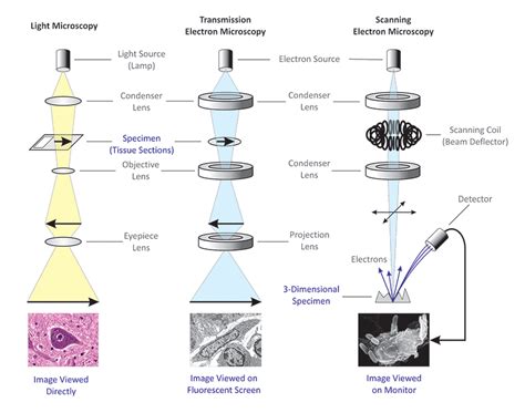
Table of Contents
Light Microscope vs. Electron Microscope: A Detailed Comparison
The world of microscopy has revolutionized our understanding of the incredibly small. From the intricacies of cellular structures to the nanoscale architecture of materials, microscopes have opened windows into realms previously invisible to the naked eye. However, not all microscopes are created equal. Two primary types dominate the field: light microscopes and electron microscopes. While both serve the purpose of magnification, they differ significantly in their mechanisms, capabilities, and applications. This comprehensive guide dives deep into the key distinctions between these two powerful tools.
Fundamental Differences: Illumination and Resolution
The most fundamental difference between light and electron microscopes lies in their method of illumination. Light microscopes use visible light to illuminate the specimen, which is then magnified using a system of lenses. Electron microscopes, on the other hand, use a beam of electrons to illuminate the specimen. This seemingly simple difference has profound implications for the resolution and capabilities of each type of microscope.
Resolution: The Key Differentiator
Resolution, the ability to distinguish between two closely spaced objects as distinct entities, is the crucial factor determining the microscope's effectiveness. The resolution of a light microscope is fundamentally limited by the wavelength of visible light. This limitation restricts the maximum resolution achievable to approximately 200 nanometers (nm). This means that objects smaller than 200 nm will appear blurred or indistinguishable.
Electron microscopes, however, achieve far higher resolution. Because the wavelength of electrons is significantly shorter than that of visible light, electron microscopes can resolve objects much smaller than light microscopes. The resolution of electron microscopes can reach down to 0.1 nm, allowing for visualization of incredibly fine details at the atomic level. This dramatic difference in resolution is a major factor in the choice between light and electron microscopy for various applications.
Magnification Capabilities
While resolution dictates the detail visible, magnification refers to the enlargement of the image. Both light and electron microscopes can achieve high magnification, but the upper limits differ. Light microscopes typically have a maximum useful magnification of around 1500x, although some specialized techniques can push this further. However, beyond this point, increasing magnification doesn't reveal further detail due to the resolution limitations mentioned earlier. The image simply becomes blurry.
Electron microscopes can achieve much higher magnifications, often exceeding 1,000,000x. This extreme magnification, coupled with their superior resolution, allows for visualization of structures at the atomic level, revealing details impossible to see with a light microscope.
Sample Preparation: A Critical Aspect
The preparation of samples for microscopy is a crucial step that significantly impacts the quality of the resulting images. Both light and electron microscopy require specific sample preparation techniques, but these differ substantially.
Light Microscopy Sample Preparation
Sample preparation for light microscopy is generally less complex and often involves simpler techniques such as:
- Staining: Using dyes to enhance contrast and highlight specific cellular structures. Different stains bind to different cellular components, allowing researchers to differentiate between various structures.
- Sectioning: Cutting thin slices of the sample to allow light to pass through. This is particularly important for thicker specimens.
- Mounting: Positioning the sample on a glass slide for observation.
Electron Microscopy Sample Preparation
Sample preparation for electron microscopy is significantly more complex and demanding. This is due to the high sensitivity of the electron beam to the sample material. Common techniques include:
- Fixation: Using chemicals to preserve the sample's structure and prevent degradation.
- Dehydration: Removing water from the sample to prevent damage from the electron beam.
- Embedding: Embedding the sample in a resin to provide structural support and allow for thin sectioning.
- Sectioning (Ultramicrotomy): Cutting extremely thin sections (typically less than 100 nm) using a specialized ultramicrotome.
- Staining (with heavy metals): Using heavy metal stains to increase electron scattering and improve contrast. These stains bind to specific cellular components, enhancing their visibility under the electron beam.
Types of Microscopy: A Closer Look
Both light and electron microscopy encompass a range of techniques, each optimized for specific applications.
Light Microscopy Techniques
- Bright-field microscopy: The most common type, where light passes directly through the specimen.
- Dark-field microscopy: Illuminates the specimen indirectly, enhancing contrast for transparent samples.
- Phase-contrast microscopy: Enhances contrast by exploiting differences in refractive index within the specimen. Excellent for observing living cells.
- Fluorescence microscopy: Uses fluorescent dyes or proteins to label specific structures within the sample. Allows for highly specific visualization and localization of molecules.
- Confocal microscopy: Uses lasers to scan the specimen, producing high-resolution optical sections. Minimizes out-of-focus blur, leading to sharper images.
Electron Microscopy Techniques
- Transmission Electron Microscopy (TEM): Electrons pass through the specimen, revealing internal structures. Provides the highest resolution among microscopy techniques.
- Scanning Electron Microscopy (SEM): Scans the specimen's surface with a focused electron beam, producing high-resolution images of surface topography. Provides 3D-like images.
- Scanning Transmission Electron Microscopy (STEM): Combines aspects of TEM and SEM, offering both high-resolution imaging and compositional analysis.
- Cryo-Electron Microscopy (Cryo-EM): Images samples in a frozen-hydrated state, preserving their native structure. Revolutionized structural biology by allowing the visualization of large, complex biomolecules.
Advantages and Disadvantages: Making the Right Choice
The choice between light and electron microscopy depends heavily on the specific application and the type of information required.
Light Microscopy Advantages:
- Relatively simple and inexpensive.
- Sample preparation is less demanding.
- Can observe living specimens.
- Versatile techniques available for different applications.
Light Microscopy Disadvantages:
- Lower resolution than electron microscopy.
- Limited magnification.
- Staining can sometimes distort the sample.
Electron Microscopy Advantages:
- Extremely high resolution.
- High magnification.
- Can reveal fine details of internal structures (TEM).
- Can provide 3D-like images of surface topography (SEM).
Electron Microscopy Disadvantages:
- Expensive and complex equipment.
- Demanding sample preparation.
- Can only observe non-living specimens (in most cases).
- Can cause damage to the sample due to the electron beam.
- Requires specialized skills and training.
Applications: Where Each Microscope Shines
The diverse applications of light and electron microscopy highlight their unique capabilities.
Light Microscopy Applications:
- Observing live cells and their processes.
- Studying cell morphology and structure.
- Identifying microorganisms.
- Analyzing tissue samples.
- Studying developmental biology.
- Medical diagnostics.
- Material science (some applications).
Electron Microscopy Applications:
- Investigating the ultrastructure of cells and organelles.
- Analyzing the structure of viruses and other nanoparticles.
- Characterizing materials at the nanoscale.
- Studying crystal structures.
- Failure analysis of materials.
- Forensic science.
- Drug discovery and development.
Conclusion: A Powerful Duo in Scientific Research
Light and electron microscopes represent two indispensable pillars of microscopy. While light microscopes offer simplicity, versatility, and the ability to study living specimens, electron microscopes provide unmatched resolution and magnification, enabling the visualization of structures at the atomic level. The choice between them depends entirely on the research question, the nature of the sample, and the level of detail required. Often, researchers use both techniques in conjunction to obtain a complete and comprehensive understanding of the subject being investigated. Their combined power continues to drive advancements in numerous fields, pushing the boundaries of scientific discovery and understanding.
Latest Posts
Latest Posts
-
What Is The Lcm Of 25 And 35
Mar 27, 2025
-
What Are All The Factors Of 11
Mar 27, 2025
-
What Is The Factors Of 25
Mar 27, 2025
-
How Much Electrons Does Sodium Have
Mar 27, 2025
-
Which Is Larger 3 8 Or 1 2
Mar 27, 2025
Related Post
Thank you for visiting our website which covers about Light Microscope And Electron Microscope Differences . We hope the information provided has been useful to you. Feel free to contact us if you have any questions or need further assistance. See you next time and don't miss to bookmark.
