Labeled Diagram Of A Prokaryotic Cell
Juapaving
Mar 16, 2025 · 6 min read
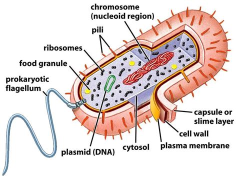
Table of Contents
Labeled Diagram of a Prokaryotic Cell: A Deep Dive into the Simplest Life Forms
Prokaryotic cells, the fundamental building blocks of bacteria and archaea, represent the simplest forms of life on Earth. Unlike their eukaryotic counterparts, they lack a membrane-bound nucleus and other complex organelles. Understanding their structure is crucial to comprehending their diverse functions and their vital roles in various ecosystems. This comprehensive guide will provide a detailed labeled diagram of a prokaryotic cell, explaining each component's function and significance.
The Basic Structure of a Prokaryotic Cell: A Visual Overview
Before diving into the specifics, let's visualize the overall structure. Imagine a simple, single-celled organism without the intricate internal compartments found in eukaryotic cells. This simplicity, however, doesn't translate to a lack of complexity in its functions. Instead, prokaryotes have evolved efficient mechanisms to perform all essential life processes within this streamlined structure.
(Here, you would insert a high-quality, labeled diagram of a prokaryotic cell. The diagram should clearly show and label all the structures discussed below. Ideally, this would be a professional-quality image or a custom-created one that is visually appealing and easy to understand. You can use tools like BioRender or draw one yourself. For the markdown version, we'll use descriptive placeholders.)
[Insert Labeled Diagram Here: This diagram should show the following structures: Plasma Membrane, Cell Wall, Capsule, Cytoplasm, Nucleoid, Ribosomes, Plasmids, Flagella, Pili, and possibly inclusion bodies.]
Key Components of a Prokaryotic Cell: A Detailed Explanation
Now let's explore each component of the prokaryotic cell in detail:
1. Plasma Membrane (Cell Membrane): The Selective Barrier
The plasma membrane, also known as the cell membrane, is a selectively permeable phospholipid bilayer surrounding the cytoplasm. This vital structure controls the passage of substances into and out of the cell, maintaining a stable internal environment. It plays a crucial role in nutrient uptake, waste excretion, and maintaining osmotic balance. The fluidity of the membrane allows for dynamic adjustments to environmental changes. The specific composition of the membrane, including the types of lipids and proteins present, varies depending on the species and environmental conditions. This variation contributes to the remarkable adaptability of prokaryotes.
2. Cell Wall: The Protective Shield
Located outside the plasma membrane, the cell wall provides structural support and protection to the cell. Unlike the cell wall of plant cells, which is made of cellulose, prokaryotic cell walls are composed primarily of peptidoglycan (in bacteria) or other similar molecules in archaea. This peptidoglycan layer is a rigid mesh-like structure that maintains the cell's shape and prevents it from bursting in hypotonic environments. The structure and composition of the cell wall are crucial in bacterial classification, with Gram-positive and Gram-negative bacteria exhibiting distinct cell wall structures. The difference in cell wall structure significantly impacts the effectiveness of antibiotics.
3. Capsule: The Protective Coat
Some prokaryotes possess a capsule, a gelatinous layer surrounding the cell wall. This external layer provides additional protection against desiccation (drying out), phagocytosis (engulfment by immune cells), and adherence to surfaces. The capsule's composition varies depending on the species and plays a role in virulence (the ability to cause disease) in pathogenic bacteria. It helps the bacteria evade the host's immune system and facilitates colonization of tissues.
4. Cytoplasm: The Cellular Matrix
The cytoplasm is the gel-like substance filling the cell interior. It's a complex mixture of water, enzymes, nutrients, wastes, and genetic material. It's the site of most metabolic reactions within the cell. Prokaryotic cytoplasm lacks the membrane-bound organelles found in eukaryotes, but it is far from empty. It's a highly organized environment containing various proteins, ribosomes, and other molecules involved in cellular processes.
5. Nucleoid: The Genetic Control Center
Unlike the membrane-bound nucleus of eukaryotes, prokaryotes have a nucleoid, a region within the cytoplasm where the genetic material (DNA) is concentrated. The prokaryotic chromosome is typically a single, circular DNA molecule that is supercoiled to fit within the nucleoid. This region is not enclosed by a membrane, unlike the eukaryotic nucleus. The nucleoid is the site of DNA replication, transcription (the process of making RNA from DNA), and other crucial genetic processes.
6. Ribosomes: Protein Synthesis Machines
Ribosomes are essential cellular structures responsible for protein synthesis. Prokaryotic ribosomes are smaller than eukaryotic ribosomes (70S versus 80S) and have a slightly different composition. They are found scattered throughout the cytoplasm and are the sites where messenger RNA (mRNA) is translated into proteins. The abundance of ribosomes reflects the high rate of protein synthesis in these rapidly growing organisms. Antibiotics like tetracycline and erythromycin specifically target prokaryotic ribosomes, inhibiting protein synthesis and ultimately killing bacterial cells.
7. Plasmids: Extrachromosomal DNA
Many prokaryotes contain plasmids, small, circular DNA molecules separate from the main chromosome. Plasmids often carry genes that provide advantages to the bacterium, such as antibiotic resistance, toxin production, or the ability to metabolize unusual substances. They can replicate independently of the chromosome and can be transferred between bacterial cells, contributing to genetic diversity and adaptation.
8. Flagella: Motility Structures
Some prokaryotes possess flagella, long, whip-like appendages used for locomotion. Bacterial flagella are different in structure and function from eukaryotic flagella. They are helical filaments that rotate to propel the cell through its environment. The number and arrangement of flagella can vary depending on the species and play a significant role in bacterial motility and chemotaxis (movement in response to chemical gradients).
9. Pili (Fimbriae): Adhesion and Conjugation
Pili are shorter, hair-like appendages found on the surface of many prokaryotes. They are involved in attachment to surfaces and other cells, playing a crucial role in biofilm formation and bacterial adherence to host tissues. A specialized type of pilus, the sex pilus, facilitates the transfer of genetic material between bacterial cells through a process called conjugation. This horizontal gene transfer plays a vital role in the spread of antibiotic resistance and other advantageous traits.
10. Inclusion Bodies: Storage Granules
Many prokaryotes contain inclusion bodies, which are storage granules that accumulate various nutrients and metabolites. These inclusions act as reserve sources of energy and building blocks for cellular components. They can store substances like glycogen (a carbohydrate), polyphosphate (a source of phosphate), or sulfur granules. The type and abundance of inclusion bodies reflect the bacterium's metabolic capabilities and the availability of nutrients in its environment.
Conclusion: The Importance of Understanding Prokaryotic Cell Structure
Understanding the structure and function of a prokaryotic cell is crucial for various fields, including medicine, biotechnology, and environmental science. The knowledge gained from studying these simple life forms has profound implications for developing new antibiotics, understanding infectious diseases, and harnessing the metabolic capabilities of microbes for biotechnological applications. The remarkable diversity and adaptability of prokaryotes, reflected in the variations in their cellular components, continue to fascinate researchers and underscore their essential roles in shaping our planet's ecosystems. Further exploration into the intricacies of these organisms will undoubtedly reveal even more about their significance in the biological world. This detailed exploration of the labeled diagram serves as a foundation for deeper understanding and further research into these fascinating microscopic entities.
Latest Posts
Latest Posts
-
Square Root Of 125 In Simplest Radical Form
Mar 16, 2025
-
Is Melting Of Wax A Physical Or Chemical Change
Mar 16, 2025
-
What Are Rows On The Periodic Table Called
Mar 16, 2025
-
Is Carbon Tetrachloride Ionic Or Covalent
Mar 16, 2025
-
Write The Formula For Sulfurous Acid
Mar 16, 2025
Related Post
Thank you for visiting our website which covers about Labeled Diagram Of A Prokaryotic Cell . We hope the information provided has been useful to you. Feel free to contact us if you have any questions or need further assistance. See you next time and don't miss to bookmark.
