Label The Diagram Of The Respiratory System
Juapaving
Mar 29, 2025 · 7 min read
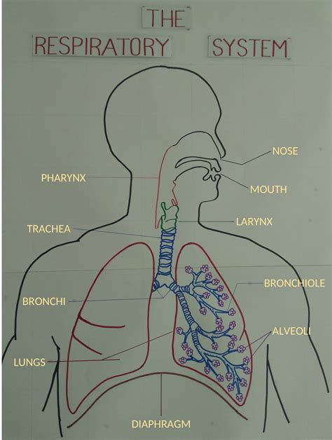
Table of Contents
Label the Diagram of the Respiratory System: A Comprehensive Guide
Understanding the respiratory system is crucial for anyone interested in biology, medicine, or simply maintaining good health. This detailed guide will not only help you label a diagram of the respiratory system but also provide a thorough understanding of its structure and function. We'll explore each component in detail, explaining its role in the complex process of breathing. By the end, you’ll be able to not only label a diagram accurately but also articulate the intricate workings of this vital system.
The Major Components of the Respiratory System
The respiratory system is responsible for the intake of oxygen and the expulsion of carbon dioxide. It’s a sophisticated network of organs and tissues working together seamlessly. The key components include:
1. The Nose and Nasal Cavity: The Gateway to Respiration
The journey of air begins in the nose and nasal cavity. The nose, the external part, acts as a protective barrier, filtering out larger particles like dust and debris. The nasal cavity, the internal space, is lined with mucous membranes which further trap and filter incoming air. These membranes also humidify and warm the air, preparing it for the delicate respiratory passages deeper within the system. The nasal conchae, bony structures within the nasal cavity, increase the surface area, enhancing the warming and humidification process. Small hairs called cilia in the nasal cavity further trap debris and move it towards the throat.
2. The Pharynx: The Crossroads of Air and Food
The pharynx, or throat, is a muscular tube that serves as a common passageway for both air and food. It’s divided into three parts: the nasopharynx, connected to the nasal cavity; the oropharynx, connected to the mouth; and the laryngopharynx, connected to the larynx. The pharynx plays a crucial role in directing air towards the larynx and food towards the esophagus. The epiglottis, a flap of cartilage, seals off the larynx during swallowing, preventing food from entering the airway. This protective mechanism is vital to prevent choking.
3. The Larynx: The Voice Box
The larynx, commonly known as the voice box, sits below the pharynx. It contains the vocal cords, which vibrate to produce sound. The larynx is composed of several cartilaginous structures, including the thyroid cartilage (Adam's apple), the cricoid cartilage, and the arytenoid cartilages. The tension and position of the vocal cords are adjusted by muscles within the larynx, enabling us to speak, sing, and whisper. The larynx also protects the lower respiratory tract by preventing the entry of food and other foreign particles.
4. The Trachea: The Windpipe
The trachea, or windpipe, is a rigid tube made of C-shaped cartilaginous rings. These rings provide structural support while allowing the trachea to expand and contract during breathing. The trachea is lined with ciliated epithelium and goblet cells, which secrete mucus to trap dust and other foreign particles. The cilia beat rhythmically to move the mucus upwards, towards the pharynx where it can be swallowed or expelled. This process, called mucociliary clearance, plays a crucial role in protecting the lungs. The trachea branches into two main bronchi, one for each lung.
5. The Bronchi and Bronchioles: Branching Pathways to the Alveoli
The main bronchi enter the lungs and further subdivide into smaller and smaller branches, forming the bronchial tree. These branches eventually lead to tiny air sacs called alveoli. The branching pattern maximizes surface area for gas exchange. The bronchi, like the trachea, are lined with ciliated epithelium and mucus-producing cells, contributing to the body’s defense mechanisms. The smallest branches, the bronchioles, are primarily composed of smooth muscle, which controls the diameter of the airways. This control is vital for regulating airflow.
6. The Lungs: The Primary Organs of Gas Exchange
The lungs are the main organs of the respiratory system. They are situated within the thoracic cavity, protected by the ribs and sternum. Each lung is divided into lobes: the right lung has three lobes (superior, middle, and inferior), and the left lung has two lobes (superior and inferior). The lungs are primarily composed of millions of tiny alveoli, the functional units of respiration. The alveoli are surrounded by a dense network of capillaries, facilitating the exchange of oxygen and carbon dioxide between the air and the bloodstream.
7. The Pleura: Protecting the Lungs
Each lung is enclosed by a double-layered membrane called the pleura. The visceral pleura is closely attached to the lung surface, while the parietal pleura lines the thoracic cavity. The space between these two layers, the pleural cavity, contains a small amount of lubricating fluid which reduces friction during breathing. This fluid also helps maintain the negative pressure within the pleural cavity, which is essential for lung expansion.
8. The Diaphragm: The Primary Muscle of Breathing
The diaphragm is a dome-shaped muscle that separates the thoracic cavity from the abdominal cavity. It plays a crucial role in breathing. During inhalation, the diaphragm contracts and flattens, increasing the volume of the thoracic cavity. This increase in volume reduces the pressure, causing air to rush into the lungs. During exhalation, the diaphragm relaxes and returns to its dome shape, decreasing the thoracic cavity volume, and forcing air out of the lungs.
9. Intercostal Muscles: Supporting Respiratory Movement
The intercostal muscles, located between the ribs, also contribute to breathing. These muscles assist in expanding and compressing the thoracic cavity during inhalation and exhalation, respectively, working in coordination with the diaphragm.
Labeling a Diagram: Step-by-Step Guide
Now that you understand the components, labeling a diagram becomes straightforward. Remember to use precise terminology. Here's a step-by-step approach:
-
Identify the Major Structures: Begin by identifying the largest and most prominent structures: the nose, nasal cavity, pharynx, larynx, trachea, lungs, and diaphragm.
-
Branching Pathways: Next, focus on the branching pattern of the airways, starting with the trachea and tracing it down to the bronchi, bronchioles, and alveoli.
-
Pleural Membranes: Locate and label the visceral and parietal pleura and the pleural cavity.
-
Detailed Structures (Optional): Depending on the complexity of the diagram, you might need to label smaller structures like the epiglottis, vocal cords, and intercostal muscles.
-
Accurate Placement: Ensure the labels are accurately placed and clearly connected to the correct structures.
-
Neatness and Clarity: Maintain neatness and clarity in your labeling, ensuring the diagram is easy to understand.
Understanding Respiratory Processes: Inhalation and Exhalation
Labeling a diagram is only the first step. A true understanding of the respiratory system involves grasping the processes of inhalation and exhalation:
Inhalation (Inspiration):
Inhalation is an active process requiring muscle contraction. The diaphragm contracts and flattens, while the intercostal muscles contract, expanding the chest cavity. This expansion reduces the pressure within the lungs, creating a pressure gradient that draws air into the respiratory system. Air flows through the nose or mouth, down the pharynx, larynx, trachea, bronchi, and finally reaches the alveoli.
Exhalation (Expiration):
Exhalation is typically a passive process. The diaphragm and intercostal muscles relax, causing the chest cavity to decrease in volume. This reduction in volume increases the pressure within the lungs, forcing air out of the respiratory system. The air travels back through the alveoli, bronchioles, bronchi, trachea, larynx, and pharynx, exiting through the nose or mouth. During strenuous activity, exhalation can become an active process, involving the contraction of abdominal muscles to further expel air.
Clinical Significance and Common Respiratory Issues
The respiratory system is susceptible to various disorders and diseases. Understanding these conditions is vital for preventative care and appropriate medical intervention. Some common respiratory issues include:
- Asthma: A chronic inflammatory condition causing airway narrowing and difficulty breathing.
- Chronic Obstructive Pulmonary Disease (COPD): A group of progressive lung diseases, including emphysema and chronic bronchitis, that obstruct airflow.
- Pneumonia: An infection of the lungs causing inflammation and fluid buildup in the alveoli.
- Lung Cancer: A serious disease characterized by uncontrolled cell growth in the lungs.
- Cystic Fibrosis: A genetic disorder affecting the respiratory system and other organs, leading to thick mucus buildup.
- Respiratory Infections (e.g., influenza, common cold): Infections that often cause temporary respiratory distress.
Conclusion
This comprehensive guide has provided you with a detailed understanding of the respiratory system, enabling you to accurately label its components on a diagram. Beyond simple labeling, we've delved into the intricate workings of inhalation and exhalation and explored some common respiratory issues. A strong understanding of the respiratory system is essential for maintaining overall health and appreciating the complexities of human biology. Remember, accurate labeling coupled with a functional understanding forms the cornerstone of comprehending this crucial system. Continue your learning journey by exploring additional resources and delving deeper into the fascinating world of human respiration.
Latest Posts
Latest Posts
-
Which Of The Following Is Not An Antibiotic
Mar 31, 2025
-
What Is Difference Between Football And Soccer
Mar 31, 2025
-
Explain The Differences Between Expressions And Equations
Mar 31, 2025
-
How Many Valence Electrons In Sr
Mar 31, 2025
-
The Last Lesson Questions And Answers
Mar 31, 2025
Related Post
Thank you for visiting our website which covers about Label The Diagram Of The Respiratory System . We hope the information provided has been useful to you. Feel free to contact us if you have any questions or need further assistance. See you next time and don't miss to bookmark.
