Label The Diagram Of The Female Reproductive System
Juapaving
Mar 19, 2025 · 7 min read
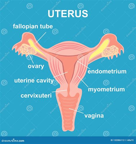
Table of Contents
Label the Diagram of the Female Reproductive System: A Comprehensive Guide
Understanding the female reproductive system is crucial for overall health and well-being. This comprehensive guide will not only help you label a diagram of the female reproductive system but also provide detailed information about the function and significance of each organ. We will cover everything from the external genitalia to the internal reproductive organs, emphasizing their intricate interplay in the process of reproduction. This article is designed to be both informative and engaging, equipping you with a solid understanding of this complex and fascinating system.
The External Genitalia: Understanding the Vulva
The external female genitalia, collectively known as the vulva, comprises several key structures:
1. Mons Pubis: The Protective Shield
The mons pubis is a fatty tissue pad located over the pubic bone. Its primary function is to protect the sensitive structures beneath it. During puberty, the mons pubis becomes covered with pubic hair, offering additional protection and playing a role in sexual attraction.
2. Labia Majora: The Outer Lips
The labia majora are two prominent folds of skin that enclose the other external genitalia. They contain sweat and oil glands, contributing to lubrication and protection. Like the mons pubis, they become covered with pubic hair during puberty. They also play a crucial role in protecting the more sensitive inner structures.
3. Labia Minora: The Inner Lips
The labia minora are two smaller folds of skin located within the labia majora. They are highly sensitive and contain numerous nerve endings. The labia minora vary significantly in size and appearance among individuals. Their primary function is to protect the clitoris and the vaginal opening.
4. Clitoris: The Center of Pleasure
The clitoris, a highly sensitive organ, is located at the upper junction of the labia minora. It's primarily composed of erectile tissue and plays a crucial role in sexual arousal and pleasure. The clitoris is rich in nerve endings, making it highly sensitive to touch and stimulation.
5. Vestibule: The Central Area
The vestibule is the area enclosed by the labia minora. It contains the openings of the vagina and urethra. The vaginal opening (introitus) may be partially covered by a thin membrane called the hymen.
6. Bartholin's Glands: Lubrication and Protection
Bartholin's glands are located on either side of the vaginal opening. They secrete a mucus-like fluid that lubricates the vagina, particularly during sexual arousal. This lubrication helps facilitate sexual intercourse and prevents discomfort.
The Internal Reproductive Organs: The Engine of Reproduction
The internal reproductive organs are responsible for producing eggs, facilitating fertilization, and nurturing the developing fetus. These organs work in a coordinated manner to achieve the complex process of reproduction.
1. Vagina: The Birth Canal and Pathway
The vagina is a muscular, elastic canal that extends from the vulva to the cervix. It serves as the birth canal during childbirth and receives the penis during sexual intercourse. The vagina also allows for the passage of menstrual flow. Its acidic environment helps to protect against infection.
2. Cervix: The Gatekeeper
The cervix is the lower, narrow part of the uterus that opens into the vagina. It plays a critical role in protecting the uterus from infection and facilitating the passage of sperm into the uterus. The cervix undergoes significant changes during menstruation and pregnancy. The opening of the cervix is known as the os.
3. Uterus: The Womb
The uterus, also known as the womb, is a pear-shaped muscular organ where a fertilized egg implants and develops into a fetus. The uterus has a thick muscular wall that allows it to expand significantly during pregnancy. The uterine lining, called the endometrium, undergoes cyclical changes throughout the menstrual cycle.
4. Fallopian Tubes: The Pathway to Fertilization
The fallopian tubes (also known as uterine tubes or oviducts) are two slender tubes that extend from the uterus to the ovaries. They transport the egg from the ovary to the uterus. Fertilization typically occurs within the fallopian tubes. The fimbriae, finger-like projections at the end of the fallopian tubes, help sweep the egg into the tube.
5. Ovaries: The Egg Factories
The ovaries are two almond-shaped organs located on either side of the uterus. They produce and release eggs (ova) as part of the menstrual cycle. The ovaries also produce hormones, such as estrogen and progesterone, which play a vital role in regulating the menstrual cycle and other bodily functions. These hormones are essential for female sexual development and reproductive health.
The Menstrual Cycle: A Monthly Rhythm
The menstrual cycle is a complex process regulated by the interplay of hormones produced by the hypothalamus, pituitary gland, and ovaries. This cyclical process prepares the body for potential pregnancy. Understanding the phases of the menstrual cycle is vital for comprehending female reproductive health.
1. Menstruation: Shedding the Lining
Menstruation is the shedding of the uterine lining (endometrium) if fertilization does not occur. This process typically lasts for 3-7 days and is accompanied by bleeding.
2. Follicular Phase: Egg Maturation
The follicular phase begins on the first day of menstruation and lasts until ovulation. During this phase, the follicle-stimulating hormone (FSH) stimulates the maturation of several follicles in the ovaries, each containing an egg.
3. Ovulation: Releasing the Egg
Ovulation is the release of a mature egg from the ovary. This usually occurs around day 14 of a 28-day cycle, although it can vary. The luteinizing hormone (LH) surge triggers ovulation.
4. Luteal Phase: Preparing for Pregnancy
The luteal phase follows ovulation and lasts until the onset of menstruation. During this phase, the ruptured follicle transforms into the corpus luteum, which produces progesterone. Progesterone prepares the uterine lining for potential implantation of a fertilized egg.
Labeling Your Diagram: A Step-by-Step Guide
Now that you have a solid understanding of the female reproductive system, let's move on to labeling a diagram. To accurately label your diagram, refer to the descriptions provided above. Here's a step-by-step guide to help you:
-
Identify the External Genitalia: Locate and label the mons pubis, labia majora, labia minora, clitoris, vestibule, and Bartholin's glands on your diagram. Pay close attention to their relative positions.
-
Identify the Internal Reproductive Organs: Locate and label the vagina, cervix, uterus, fallopian tubes, and ovaries. Understand their interconnectedness.
-
Add Details (Optional): For a more comprehensive diagram, you could add details such as the fimbriae of the fallopian tubes, the endometrium of the uterus, and the os of the cervix.
By carefully following these steps and referring to the descriptions provided, you will be able to accurately label a diagram of the female reproductive system.
Beyond Labeling: Understanding Reproductive Health
Understanding the female reproductive system goes beyond simply labeling a diagram. It's crucial to be aware of potential health issues and seek appropriate medical care when necessary. Some common concerns include:
- Menstrual irregularities: Changes in the frequency, duration, or heaviness of menstrual periods.
- Pelvic inflammatory disease (PID): An infection of the female reproductive organs.
- Endometriosis: A condition where tissue similar to the uterine lining grows outside the uterus.
- Ovarian cysts: Fluid-filled sacs on the ovaries.
- Uterine fibroids: Benign tumors in the uterus.
- Cervical cancer: Cancer of the cervix.
- Ovarian cancer: Cancer of the ovaries.
Regular check-ups with a gynecologist are essential for maintaining reproductive health and detecting potential problems early. Early detection significantly improves the chances of successful treatment.
Conclusion: A Journey of Understanding
This comprehensive guide has provided you with a detailed understanding of the female reproductive system, equipping you to accurately label a diagram and grasp the intricate workings of this vital system. Remember that this information is for educational purposes only and should not be considered medical advice. Always consult with a healthcare professional for any health concerns. By understanding the female reproductive system, we empower ourselves to make informed decisions about our health and well-being. The knowledge gained here provides a foundation for a deeper appreciation of the complexity and beauty of the human body. Continuously seeking knowledge and engaging in open discussions about reproductive health are crucial steps in maintaining a healthy and fulfilling life.
Latest Posts
Latest Posts
-
What Is The Velocity Of Light In A Vacuum
Mar 20, 2025
-
Least Common Multiple Of 4 5 And 6
Mar 20, 2025
-
The Ability To Do Work Is Called
Mar 20, 2025
-
How Many Lines Of Symmetry Has A Pentagon
Mar 20, 2025
-
Levels Of Organization In Multicellular Organisms
Mar 20, 2025
Related Post
Thank you for visiting our website which covers about Label The Diagram Of The Female Reproductive System . We hope the information provided has been useful to you. Feel free to contact us if you have any questions or need further assistance. See you next time and don't miss to bookmark.
