How Does An Electron Microscope Differ From A Light Microscope
Juapaving
Mar 31, 2025 · 5 min read

Table of Contents
How Does an Electron Microscope Differ From a Light Microscope?
The world of microscopy has revolutionized our understanding of the incredibly small. From the intricate details of a single cell to the atomic structure of materials, microscopy allows us to visualize and analyze the unseen. Two prominent techniques dominate this field: light microscopy and electron microscopy. While both aim to magnify images beyond the capacity of the naked eye, their underlying principles, capabilities, and applications differ significantly. This article delves into the key distinctions between these two powerful tools, highlighting their strengths and limitations.
Fundamental Differences: Light vs. Electron Beams
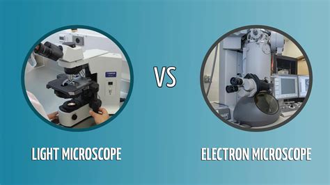
The most fundamental difference lies in the type of illumination used. Light microscopes utilize visible light, focused using lenses, to illuminate and magnify a specimen. This relatively simple approach has been around for centuries and allows for real-time observation of living samples. However, the resolving power of light microscopy is limited by the wavelength of light.
Electron microscopes, on the other hand, employ a beam of electrons instead of light. Electrons have a significantly shorter wavelength than visible light, enabling vastly higher resolution. This allows electron microscopy to reveal structures far smaller than those visible under even the most powerful light microscopes. The focusing of the electron beam is achieved using electromagnetic lenses rather than glass lenses.
Wavelength and Resolution: The Key Differentiator
The resolving power, or the ability to distinguish between two closely spaced objects, is directly related to the wavelength of the illuminating source. Since electrons have a much shorter wavelength than photons (light particles), electron microscopes achieve significantly higher resolution, typically in the nanometer range, compared to the micrometer range achievable with light microscopy. This means electron microscopes can visualize structures hundreds of times smaller than light microscopes.
- Light Microscopy Resolution: ~200 nm (limited by the diffraction of light)
- Electron Microscopy Resolution: ~0.1 nm (depending on the type of electron microscope)
Types of Microscopes and Their Applications
Both light and electron microscopy encompass various types, each optimized for specific applications:
Light Microscopy Types:
- Bright-field microscopy: The most common type, where light passes directly through the specimen. Simple and versatile, but often requires staining to enhance contrast. Suitable for observing stained cells, tissues, and some microorganisms.
- Dark-field microscopy: Light is scattered by the specimen, creating a bright image against a dark background. Ideal for observing unstained, transparent specimens like live bacteria.
- Phase-contrast microscopy: Exploits differences in refractive index to visualize transparent specimens without staining. Excellent for observing living cells and their internal structures.
- Fluorescence microscopy: Uses fluorescent dyes or proteins to label specific structures within a sample. Allows for the visualization of specific molecules and processes within cells. Widely used in cell biology and medical diagnostics.
- Confocal microscopy: A sophisticated technique that uses lasers to scan a specimen, creating incredibly sharp, three-dimensional images. Excellent for visualizing thick specimens and resolving complex structures.
Electron Microscopy Types:
- Transmission Electron Microscopy (TEM): Electrons pass through a very thin specimen, creating a high-resolution image based on electron scattering and absorption. Provides detailed information about the internal structure of cells, materials, and even individual atoms.
- Scanning Electron Microscopy (SEM): Electrons scan the surface of a specimen, producing a detailed three-dimensional image based on the detection of scattered electrons. Ideal for visualizing surface features and topography, providing high-resolution images of complex structures.
- Scanning Transmission Electron Microscopy (STEM): A hybrid technique combining aspects of TEM and SEM. Provides both high-resolution imaging of internal structures and surface details.
Sample Preparation: A Crucial Difference
Sample preparation is a critical aspect of microscopy, and the methods employed differ greatly between light and electron microscopy.
Light microscopy typically requires less rigorous preparation. Specimens can often be simply mounted on a glass slide, possibly stained to enhance contrast. Living samples can also be directly observed.
Electron microscopy, on the other hand, demands much more meticulous preparation. Specimens must be carefully fixed, dehydrated, and embedded in resin before being sectioned into extremely thin slices (for TEM) or coated with a conductive material (for SEM). This intricate process can be time-consuming and may introduce artifacts that affect the final image. Furthermore, electron microscopy typically requires samples to be in a vacuum, rendering it unsuitable for observing live specimens.
Cost and Complexity: A Significant Factor
Light microscopes are generally less expensive and easier to operate than electron microscopes. Their relative simplicity makes them accessible to a wider range of users and educational settings.
Electron microscopes, however, are significantly more expensive to purchase, maintain, and operate. They require specialized training and a dedicated facility with appropriate infrastructure, including vacuum pumps and high-voltage power supplies. This high cost limits their accessibility to specialized research laboratories and institutions.
Image Interpretation and Analysis: Different Approaches
Interpreting images from light and electron microscopy also requires different approaches. Light microscopy images are often relatively straightforward to interpret, although specialized techniques like fluorescence microscopy may require additional knowledge of the specific fluorophores used.
Electron microscopy images, especially those from TEM, can be more complex to interpret, requiring understanding of diffraction patterns, contrast mechanisms, and the effects of sample preparation. Image analysis software is often employed to enhance the images and extract quantitative data.
Specific Applications: Where Each Technique Excels
The choice between light and electron microscopy depends heavily on the specific application and the information required.
Light microscopy is well-suited for:
- Observing living cells and their dynamic processes.
- Studying larger structures like tissues and organs.
- Performing routine clinical diagnostics.
- Educational purposes, providing a relatively simple and accessible method for visualizing microscopic structures.
Electron microscopy is essential for:
- Visualizing structures at the nanometer scale.
- Studying the internal structure of cells and organelles in great detail.
- Analyzing materials at the atomic level.
- Imaging the surface topography of specimens with exceptional clarity.
- Investigating viruses and other nanostructures.
Conclusion: Complementary Techniques
Light and electron microscopy are not mutually exclusive; they are often complementary techniques. Light microscopy can provide a broader overview of a specimen, guiding the selection of regions for more detailed investigation using electron microscopy. The combination of these techniques offers the most complete understanding of the microscopic world. Each technique excels in specific areas, dictated by resolution needs, sample characteristics, and available resources. The ongoing advancements in both fields continue to push the boundaries of our understanding of the microcosm, uncovering new insights into the structure and function of life and materials.
Latest Posts
Latest Posts
-
Three Or More Points That Lie In The Same Line
Apr 02, 2025
-
Whats A Shape With 8 Vertices And 6 Faces
Apr 02, 2025
-
6 Quarts Of Water To Cups
Apr 02, 2025
-
What Is The Greatest Common Factor Of 49
Apr 02, 2025
-
The First Organisms On Earth Were
Apr 02, 2025
Related Post
Thank you for visiting our website which covers about How Does An Electron Microscope Differ From A Light Microscope . We hope the information provided has been useful to you. Feel free to contact us if you have any questions or need further assistance. See you next time and don't miss to bookmark.
