Diagram Of Digestive System Of Frog
Juapaving
Mar 25, 2025 · 5 min read
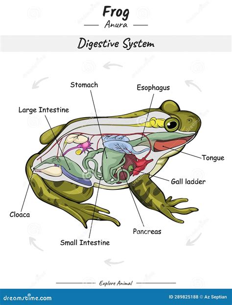
Table of Contents
Diagram of the Frog Digestive System: A Comprehensive Guide
The frog, a fascinating amphibian, possesses a digestive system remarkably adapted to its carnivorous diet and amphibious lifestyle. Understanding its anatomy provides valuable insights into the broader principles of vertebrate digestion and the specific adaptations required for survival in diverse environments. This comprehensive guide will explore the frog digestive system in detail, using diagrams and explanations to clarify its structure and function.
The Frog's Digestive Tract: A Journey Through the System
The frog's digestive system is a continuous tube, beginning at the mouth and ending at the cloaca. This journey involves a series of specialized organs working in concert to break down food, absorb nutrients, and eliminate waste. Let's trace this path step by step:
1. Buccal Cavity (Mouth): The Beginning of Digestion
The journey begins in the buccal cavity, or mouth. Frogs lack chewing teeth, instead using their sticky tongue to capture prey. The tongue, attached to the front of the mouth, is rapidly projected to snatch insects and other small animals. Within the buccal cavity, salivary glands secrete mucus, which lubricates the food, making it easier to swallow. While the saliva doesn't contain digestive enzymes in the same way as mammals, the mucus assists in the initial stages of food breakdown. Small, backward-pointing teeth line the jaws, holding the prey firmly in place while it's swallowed whole.
2. Oesophagus: Transporting Food to the Stomach
Once secured, the prey is swallowed whole. This leads to the oesophagus, a short, muscular tube connecting the buccal cavity to the stomach. The oesophagus's peristaltic contractions—rhythmic wave-like movements—push the food down to the stomach. These contractions are crucial for transporting food efficiently, even against gravity.
3. Stomach: Chemical Breakdown Begins
The stomach, a J-shaped organ, is the site of significant chemical digestion. Gastric glands within the stomach lining secrete gastric juice, a mixture of hydrochloric acid (HCl) and pepsinogen. HCl creates a highly acidic environment, killing ingested bacteria and activating pepsinogen into pepsin, a protease enzyme that begins breaking down proteins. The stomach's muscular walls churn the food, mixing it thoroughly with gastric juice to enhance digestion. This process produces a semi-fluid mixture called chyme.
4. Small Intestine: Nutrient Absorption
The chyme then passes into the small intestine, a long, coiled tube responsible for the majority of nutrient absorption. The small intestine consists of three parts: the duodenum, jejunum, and ileum. The duodenum receives secretions from the pancreas and liver.
-
Pancreas: The pancreas secretes pancreatic juice, containing various digestive enzymes including trypsin (another protein-digesting enzyme), amylase (carbohydrate digestion), and lipase (fat digestion). This ensures the complete breakdown of proteins, carbohydrates and fats, allowing for efficient absorption.
-
Liver: The liver produces bile, stored in the gallbladder. Bile emulsifies fats, breaking them down into smaller droplets, increasing the surface area for lipase to act upon. This process enhances fat digestion and absorption.
The jejunum and ileum have a highly folded inner surface, dramatically increasing surface area for nutrient absorption. Nutrients, broken down into their simplest forms (amino acids, glucose, fatty acids, etc.), are absorbed through the intestinal wall and enter the bloodstream, transporting them throughout the body.
5. Large Intestine: Water Absorption and Waste Elimination
Following nutrient absorption in the small intestine, the indigestible remains enter the large intestine, a shorter, wider tube. The large intestine's primary function is to absorb water from the remaining undigested material, forming solid waste. This waste, along with any remaining undigested material, is then passed to the cloaca.
6. Cloaca: The Final Destination
The cloaca is a common chamber where the digestive, urinary, and reproductive tracts converge. Waste products from the digestive system (faeces), urine, and reproductive products are expelled from the body through the cloaca. This is a characteristic feature of amphibians, reptiles, and birds.
Diagrammatic Representation of the Frog Digestive System
(Imagine a diagram here showing the mouth, oesophagus, stomach, small intestine, large intestine, pancreas, liver, gallbladder and cloaca, clearly labelled and demonstrating the flow of food.)
The diagram should visually represent the connections and relative sizes of the organs, enhancing understanding of the system's overall structure. A well-labeled diagram would highlight the key features and functions of each organ, making it an effective learning tool.
Adaptations of the Frog Digestive System
Several adaptations in the frog's digestive system reflect its lifestyle and diet:
- Sticky Tongue: This allows for efficient capture of fast-moving prey.
- Short Oesophagus: Enables rapid transport of food to the stomach.
- Acidic Stomach: Kills bacteria ingested with prey, initiating protein digestion.
- Efficient Small Intestine: Facilitates complete nutrient absorption, crucial for meeting energy demands.
- Cloaca: A common exit point for various bodily waste products, maximizing efficiency.
These adaptations highlight the evolutionary pressures shaping the structure and function of the frog's digestive system.
Comparing Frog and Mammalian Digestive Systems
While sharing some basic similarities, frog and mammalian digestive systems differ significantly. Mammals possess more specialized teeth for chewing, increasing surface area for enzymatic action. Their digestive tracts are often longer, reflecting a more varied diet. The absence of a cloaca in mammals is a notable difference, with separate openings for the urinary, digestive, and reproductive systems. The key differences stem from their contrasting diets and environmental adaptations.
Conclusion: The Importance of Understanding the Frog Digestive System
Understanding the frog digestive system provides a foundational understanding of digestive physiology in vertebrates. The system's unique adaptations showcase how organisms evolve to meet specific ecological demands. By studying the frog's digestive system, we gain insights into broader biological principles, contributing to a deeper appreciation of the intricate mechanisms that sustain life. Furthermore, the frog's digestive system serves as a valuable model for research in various areas of biology, from comparative anatomy to the study of digestive enzymes. The simplicity and accessibility of the frog's system makes it an ideal subject for educational purposes, furthering our understanding of life's fundamental processes. This comprehensive analysis, coupled with a well-illustrated diagram, provides a robust understanding of the frog’s fascinating digestive system.
Latest Posts
Latest Posts
-
State Newtons Law Of Universal Gravitation In Words
Mar 28, 2025
-
Is 6 A Factor Of 84
Mar 28, 2025
-
What Is The Lcm Of 2 And 11
Mar 28, 2025
-
In The Burning Of Methane What Are The Reactants
Mar 28, 2025
-
What Is The Difference Between Methanol And Ethanol Fuels
Mar 28, 2025
Related Post
Thank you for visiting our website which covers about Diagram Of Digestive System Of Frog . We hope the information provided has been useful to you. Feel free to contact us if you have any questions or need further assistance. See you next time and don't miss to bookmark.
