A Sarcomere Is A Regions Between Two
Juapaving
Apr 02, 2025 · 6 min read

Table of Contents
A Sarcomere: The Region Between Two Z-Lines – A Deep Dive into Muscle Contraction
The human body is a marvel of engineering, capable of feats of strength, endurance, and precision. At the heart of this capability lies the muscle, and within the muscle, the microscopic powerhouse responsible for all movement: the sarcomere. This article will delve deep into the fascinating world of the sarcomere, exploring its structure, function, and the intricate mechanisms driving muscle contraction. We'll unravel the complexities of this fundamental unit of muscle, explaining its significance in everything from a gentle hand movement to a powerful athletic performance.
Understanding the Sarcomere: The Basic Unit of Muscle Contraction
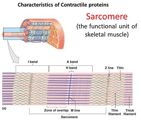
A sarcomere is defined as the region between two Z-lines (or Z-discs). These Z-lines are dense protein structures that serve as the anchoring points for the contractile filaments within the muscle fiber. Imagine a sarcomere as a tiny, highly organized machine packed with proteins, meticulously arranged to perform a single, crucial task: muscle contraction. Understanding the sarcomere's structure is essential to understanding how muscles work.
The Key Players: Actin and Myosin Filaments
The sarcomere's contractile machinery primarily consists of two types of protein filaments:
-
Actin filaments (thin filaments): These are long, thin strands composed of actin protein molecules. They are anchored to the Z-lines and extend towards the center of the sarcomere. Think of these as the "rails" along which the other filament moves.
-
Myosin filaments (thick filaments): These are thicker filaments composed of myosin protein molecules. They are located in the center of the sarcomere, overlapping with the actin filaments. These are the "motors" driving the contraction. Each myosin molecule has a head that can bind to actin and generate force.
The precise arrangement and interaction of these filaments are crucial for the sarcomere's function. The overlap between actin and myosin filaments is what dictates the length and strength of the sarcomere and ultimately, the muscle.
The Sliding Filament Theory: How Muscles Contract
The mechanism by which muscles contract is known as the sliding filament theory. This theory elegantly explains how the interaction between actin and myosin filaments leads to muscle shortening. Here's a step-by-step breakdown:
-
Calcium Ion Release: Muscle contraction is initiated by a nerve impulse that triggers the release of calcium ions (Ca²⁺) into the sarcoplasm (the cytoplasm of the muscle cell).
-
Calcium Binding to Troponin: These calcium ions bind to a protein complex called troponin, which is located on the actin filament.
-
Troponin-Tropomyosin Shift: This binding causes a conformational change in troponin, shifting another protein called tropomyosin. Tropomyosin usually blocks the myosin-binding sites on the actin filament. This shift exposes these binding sites.
-
Cross-Bridge Formation: Now, the myosin heads can bind to the exposed binding sites on the actin filament, forming what are called cross-bridges.
-
Power Stroke: The myosin heads then undergo a conformational change, pivoting and pulling the actin filament towards the center of the sarcomere. This is the power stroke, generating the force of muscle contraction.
-
ATP Hydrolysis and Detachment: The energy for this power stroke comes from the hydrolysis of ATP (adenosine triphosphate). After the power stroke, ATP binds to the myosin head, causing it to detach from the actin filament.
-
Cycle Repetition: The cycle then repeats itself, with myosin heads continuously binding, pulling, detaching, and re-binding, resulting in the sliding of actin filaments over myosin filaments. This process continues as long as calcium ions are present and ATP is available.
-
Sarcomere Shortening: As the actin filaments slide over the myosin filaments, the sarcomere shortens, leading to muscle contraction. The Z-lines move closer together, reflecting this shortening.
Sarcomere Structure in Detail: Beyond the Basics
While the simplified explanation above captures the essence of muscle contraction, the sarcomere's structure possesses remarkable intricacy. Let's explore some of its key components in more detail:
The Z-Line: The Anchoring Point
The Z-line (or Z-disc) is a protein structure that serves as the attachment point for the thin filaments (actin). It’s a complex structure containing various proteins, including α-actinin, which helps to anchor the actin filaments and maintain the structural integrity of the sarcomere. The Z-line's position and stability are crucial for proper muscle function.
The I-Band: The Light Band
The I-band is the region of the sarcomere containing only actin filaments. It appears lighter under a microscope because it lacks the dense myosin filaments. The I-band shortens during muscle contraction as the actin filaments slide towards the center of the sarcomere.
The A-Band: The Dark Band
The A-band is the region of the sarcomere containing both actin and myosin filaments. It appears darker under a microscope because of the dense packing of filaments. The A-band's length remains relatively constant during muscle contraction, even though the actin and myosin overlap changes.
The H-Zone: The Myosin-Only Zone
The H-zone is the region within the A-band containing only myosin filaments. It appears lighter than the rest of the A-band because it lacks the overlap with actin filaments. During muscle contraction, the H-zone shortens as the actin filaments slide inwards.
The M-Line: The Middle Line
The M-line is located in the center of the sarcomere, within the H-zone. It's a protein structure that holds the myosin filaments in place and helps to maintain the sarcomere's organization. Proteins like myomesin and M-protein contribute to the M-line's structural integrity.
Types of Muscle Fibers and Sarcomere Characteristics
Different types of muscle fibers have varying sarcomere structures and contractile properties:
-
Type I (Slow-twitch) fibers: These fibers are characterized by their slow contraction speed and high endurance. They generally have a higher density of mitochondria (the energy powerhouses of the cell) and a greater capacity for oxidative metabolism. Their sarcomeres may show slightly different arrangements of proteins compared to fast-twitch fibers.
-
Type II (Fast-twitch) fibers: These fibers are characterized by their fast contraction speed and high power output but lower endurance. They rely more on glycolytic metabolism (anaerobic energy production). Their sarcomere structure might exhibit variations in filament length and overlap, contributing to their speed and power.
Sarcomere Dysfunction and Muscle Diseases
Disruptions in the sarcomere's structure or function can lead to a variety of muscle diseases. These diseases often involve mutations in the genes encoding the sarcomeric proteins, leading to weakened or dysfunctional muscles. Examples include:
-
Muscular dystrophy: A group of inherited diseases characterized by progressive muscle degeneration and weakness.
-
Cardiomyopathies: Diseases affecting the heart muscle, often involving abnormalities in sarcomere structure and function.
-
Myopathies: A broad category of muscle diseases affecting muscle function and structure, often involving sarcomeric abnormalities.
Conclusion: The Sarcomere's Crucial Role
The sarcomere, the region between two Z-lines, stands as a testament to the intricate elegance of biological systems. Its precise architecture and the precise interplay of its protein components enable muscle contraction, the fundamental process underpinning all forms of movement. From the smallest twitch to the most powerful exertion, the sarcomere plays an indispensable role. Understanding its structure and function is crucial not only for comprehending basic physiology but also for developing effective treatments for muscle diseases that affect millions worldwide. Further research into sarcomere biology continues to unveil new insights into muscle function and dysfunction, paving the way for advancements in healthcare and athletic performance enhancement. The microscopic world within our muscles holds secrets yet to be revealed, further emphasizing the remarkable complexity and ingenuity of the human body.
Latest Posts
Latest Posts
-
Good Words To Describe Your Mother
Apr 03, 2025
-
How Many Bones Does Shark Have
Apr 03, 2025
-
Ca Oh 2 Acid Or Base
Apr 03, 2025
-
Quadrilateral With One Pair Of Parallel Sides
Apr 03, 2025
-
Any Change In Velocity Is Called
Apr 03, 2025
Related Post
Thank you for visiting our website which covers about A Sarcomere Is A Regions Between Two . We hope the information provided has been useful to you. Feel free to contact us if you have any questions or need further assistance. See you next time and don't miss to bookmark.
