A Compound Microscope Is A Type Of Optical Microscope.
Juapaving
Apr 05, 2025 · 6 min read

Table of Contents
A Compound Microscope: Delving Deep into the World of Optical Microscopy
The compound microscope, a cornerstone of scientific exploration, stands as a testament to human ingenuity in unraveling the intricacies of the microscopic world. This powerful optical instrument, capable of magnifying images far beyond the capabilities of the human eye, has revolutionized fields ranging from biology and medicine to materials science and engineering. This article delves into the fascinating world of compound microscopy, exploring its components, principles, applications, and the ongoing advancements shaping its future.
Understanding the Fundamentals of Compound Microscopy
Unlike simple microscopes, which use a single lens for magnification, compound microscopes employ a system of two lenses: the objective lens and the eyepiece lens (ocular lens). This dual-lens system allows for significantly higher magnification and resolution, revealing intricate details invisible to the naked eye or even a simple microscope.
The Objective Lens: The First Stage of Magnification
The objective lens, located closest to the specimen, is responsible for the primary magnification. It creates a real, inverted, and magnified image of the specimen. Compound microscopes typically offer a revolving nosepiece housing several objective lenses with varying magnification powers, commonly ranging from 4x (low power) to 100x (oil immersion high power). Each objective lens is meticulously designed and constructed to minimize aberrations (distortions) and maximize image clarity. High-quality objective lenses are crucial for achieving optimal resolution and image fidelity.
The Eyepiece Lens: The Final Image Formation
The eyepiece lens (ocular lens) further magnifies the already magnified image produced by the objective lens. It takes the real image from the objective lens and produces a virtual, magnified, and inverted image that the observer views directly. Eyepieces typically have magnifications of 10x, although other magnifications are available. The total magnification of the compound microscope is calculated by multiplying the magnification of the objective lens by the magnification of the eyepiece lens. For example, a 40x objective lens used with a 10x eyepiece yields a total magnification of 400x.
Resolution: Beyond Magnification
While magnification is crucial, it's equally important to understand the concept of resolution, which refers to the ability of a microscope to distinguish between two closely spaced objects as distinct entities. High magnification without high resolution results in a blurry, indistinct image. The resolution of a compound microscope is largely determined by the numerical aperture (NA) of the objective lens and the wavelength of light used. A higher NA and shorter wavelength result in better resolution.
Key Components of a Compound Microscope
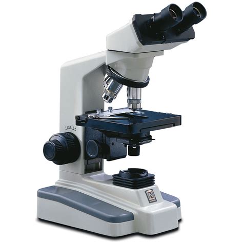
A typical compound microscope comprises several essential components working in concert to produce a magnified image:
- Base: Provides structural support for the microscope.
- Arm: Connects the base to the body tube, providing a stable handle for carrying the microscope.
- Stage: A platform where the specimen slide is placed. It often includes mechanical stage controls for precise specimen movement.
- Stage Clips: Secure the specimen slide in place on the stage.
- Illumination System: Provides the light source for illuminating the specimen. This can include a built-in lamp or an external light source. Köhler illumination is a technique often used to ensure even and optimal illumination across the specimen.
- Condenser: Focuses the light onto the specimen, controlling the intensity and distribution of light. Adjusting the condenser is crucial for achieving optimal image contrast and resolution.
- Iris Diaphragm: Controls the amount of light passing through the condenser, influencing contrast and depth of field.
- Coarse Adjustment Knob: Moves the stage up and down in large increments for initial focusing.
- Fine Adjustment Knob: Makes smaller, more precise adjustments to the stage for fine focusing.
- Body Tube: Connects the objective lens to the eyepiece lens, maintaining the correct optical path.
- Revolving Nosepiece: Holds multiple objective lenses, allowing for quick and easy changes in magnification.
- Objective Lenses: Magnify the specimen, providing different levels of magnification.
- Eyepiece Lens (Ocular Lens): Further magnifies the image from the objective lens for viewing.
Types of Compound Microscopes
While the basic principles remain the same, variations exist in compound microscopes designed for specific applications:
- Brightfield Microscopes: These are the most common type, where light passes directly through the specimen. Staining is often required to enhance contrast.
- Darkfield Microscopes: In this type, only scattered light from the specimen enters the objective lens, making the specimen appear bright against a dark background. This is useful for observing unstained specimens.
- Phase-Contrast Microscopes: These enhance contrast by exploiting differences in the refractive indices of various parts of the specimen, allowing for the visualization of transparent structures.
- Fluorescence Microscopes: These use fluorescent dyes or proteins to label specific structures within the specimen, allowing for highly specific and sensitive imaging. Confocal microscopy is a sophisticated form of fluorescence microscopy that eliminates out-of-focus light, producing high-resolution images.
- Polarizing Microscopes: These utilize polarized light to analyze the optical properties of materials, useful in identifying crystalline structures and birefringent materials.
Preparing Specimens for Compound Microscopy
Proper specimen preparation is critical for obtaining high-quality images. Methods vary depending on the type of specimen and the desired information:
- Smear Preparations: Used for liquid samples like blood or bacteria.
- Squash Preparations: Gentle pressing of the specimen onto a slide.
- Sectioning: Thin slices of tissues or materials are prepared using microtomes.
- Staining Techniques: A wide range of dyes are used to enhance contrast and highlight specific structures. Gram staining, for example, is crucial in microbiology.
- Mounting Media: A transparent medium is used to protect and preserve the specimen on the slide.
Applications of Compound Microscopes
The applications of compound microscopes are extensive and span across numerous disciplines:
- Biology and Medicine: Observing cells, tissues, microorganisms, and pathogens. Critical for diagnosis, research, and education in fields like histology, pathology, microbiology, and cytology.
- Materials Science: Analyzing the microstructure of materials, identifying defects, and studying material properties at the microscopic level. Essential in metallurgy, ceramics, and polymer science.
- Forensic Science: Examining trace evidence, fibers, hairs, and other forensic samples.
- Environmental Science: Analyzing water samples for microorganisms and pollutants.
- Education: A fundamental tool in science education, enabling students to explore the microscopic world.
Advances in Compound Microscopy
Technological advancements continue to improve the capabilities of compound microscopes:
- Digital Microscopy: Integrating digital cameras allows for image capture, processing, and storage. Software can enhance image quality and provide quantitative analysis.
- Automated Microscopy: Automating functions like focusing, stage movement, and image acquisition increases efficiency and throughput, particularly in high-throughput screening applications.
- Super-Resolution Microscopy: Techniques like PALM (Photoactivated Localization Microscopy) and STORM (Stochastic Optical Reconstruction Microscopy) surpass the diffraction limit of light, enabling the visualization of structures smaller than the wavelength of light.
- Light Sheet Microscopy: This technique illuminates the specimen with a thin sheet of light, reducing photobleaching and phototoxicity, allowing for the visualization of three-dimensional structures with high speed and resolution.
Conclusion: The Enduring Power of the Compound Microscope
The compound microscope remains a vital instrument in scientific research and technological advancement. Its ability to reveal the intricate details of the microscopic world continues to drive innovation across diverse fields. From its humble beginnings to its sophisticated modern iterations, the compound microscope has undoubtedly shaped our understanding of the universe at its smallest scales, and its future holds the promise of even more groundbreaking discoveries. The ongoing development of new techniques and technologies ensures that the compound microscope will continue to play a critical role in scientific exploration for years to come. Its enduring power lies not only in its ability to magnify images but also in its capacity to illuminate our understanding of the world around us.
Latest Posts
Latest Posts
-
Least Common Multiple 4 And 8
Apr 05, 2025
-
What Is The Atomic Mass Of Magnesium
Apr 05, 2025
-
A Pectoral Girdle Consists Of Two Bones The And The
Apr 05, 2025
-
How Many Feet Is 26 Inches
Apr 05, 2025
-
Actin And Myosin Are What Type Of Biological Molecule
Apr 05, 2025
Related Post
Thank you for visiting our website which covers about A Compound Microscope Is A Type Of Optical Microscope. . We hope the information provided has been useful to you. Feel free to contact us if you have any questions or need further assistance. See you next time and don't miss to bookmark.
