A Pectoral Girdle Consists Of Two Bones The And The
Juapaving
Apr 05, 2025 · 7 min read

Table of Contents
A Pectoral Girdle Consists of Two Bones: The Clavicle and the Scapula
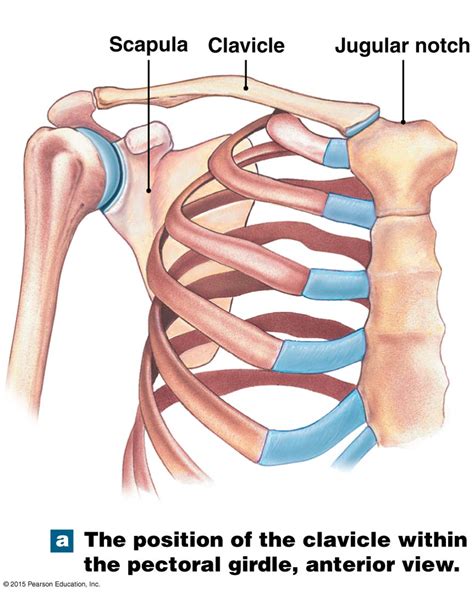
The human pectoral girdle, also known as the shoulder girdle, is a complex anatomical structure responsible for connecting the upper limbs to the axial skeleton. Unlike the more stable pelvic girdle, the pectoral girdle prioritizes mobility over stability, allowing for a remarkable range of arm movements. This remarkable flexibility comes at the cost of inherent instability, making it prone to injuries like dislocations. Understanding the anatomy of the pectoral girdle, specifically the two bones that constitute it—the clavicle and the scapula—is crucial for appreciating its function and vulnerability.
The Clavicle: The Collarbone's Vital Role
The clavicle, commonly known as the collarbone, is a long, S-shaped bone situated at the base of the neck. It's easily palpable just beneath the skin, forming a prominent landmark on the anterior aspect of the shoulder. Its unique shape and position are critical to its function.
Anatomical Features of the Clavicle:
- Sternal End: The medial, or inner, end of the clavicle articulates with the manubrium of the sternum (breastbone) at the sternoclavicular joint (SC joint). This joint is the only bony connection between the upper limb and the axial skeleton.
- Acromial End: The lateral, or outer, end of the clavicle articulates with the acromion process of the scapula, forming the acromioclavicular joint (AC joint). This joint allows for a gliding motion between the clavicle and scapula.
- Conoid Tubercle: A roughened area on the inferior surface of the acromial end, serving as an attachment point for ligaments.
- Costal Groove: A shallow groove located on the inferior surface, providing passage for the subclavian vein and the subclavian artery.
Functional Significance of the Clavicle:
The clavicle plays several crucial roles:
- Transmission of Forces: It acts as a strut, transferring forces from the upper limb to the axial skeleton. This is vital for activities involving pushing, pulling, and weight-bearing. Imagine the impact on your body if this force wasn't effectively transferred!
- Shoulder Stability: Although the shoulder joint itself is inherently unstable, the clavicle contributes to overall shoulder stability by providing a rigid framework and acting as a lever arm for muscles.
- Range of Motion: The clavicle's unique articulation with both the sternum and scapula allows for a wide range of shoulder movements, including flexion, extension, abduction, adduction, internal rotation, and external rotation. Without the clavicle's contribution, the range of motion would be significantly reduced.
- Protection of Neurovascular Structures: The clavicle shields underlying neurovascular structures, including the brachial plexus and subclavian vessels, from trauma.
The Scapula: The Shoulder Blade's Complex Anatomy
The scapula, commonly known as the shoulder blade, is a large, flat triangular bone situated on the posterior aspect of the thorax. Unlike the clavicle, the scapula doesn't directly articulate with the axial skeleton. Instead, it's connected through the clavicle and a complex network of muscles and ligaments.
Anatomical Features of the Scapula:
- Acromion Process: A bony projection extending laterally from the superior angle, forming the lateral end of the spine of the scapula. It articulates with the clavicle at the AC joint.
- Coracoid Process: A hook-like projection extending anteriorly from the superior border. It serves as an attachment point for various muscles.
- Glenoid Cavity (Fossa): A shallow, pear-shaped depression on the lateral aspect of the scapula. It articulates with the head of the humerus, forming the glenohumeral joint (shoulder joint). The shallowness of this cavity contributes to the shoulder's mobility and instability.
- Spine of the Scapula: A prominent ridge extending diagonally across the posterior surface, providing attachment sites for muscles.
- Supraspinous Fossa: The fossa above the spine, housing the supraspinatus muscle.
- Infraspinous Fossa: The fossa below the spine, housing the infraspinatus muscle.
- Subscapular Fossa: The concave surface on the anterior aspect of the scapula, housing the subscapularis muscle.
- Superior, Medial, and Inferior Borders: These borders define the shape and dimensions of the scapula.
- Superior and Inferior Angles: These bony landmarks represent the corners of the scapula.
Functional Significance of the Scapula:
The scapula's unique anatomy contributes significantly to shoulder function:
- Scapulohumeral Rhythm: The scapula plays a crucial role in scapulohumeral rhythm, the coordinated movement between the scapula and humerus that optimizes shoulder range of motion. This intricate dance of bones and muscles allows for a wide variety of movements.
- Glenohumeral Joint Stability: Although not directly involved in the glenohumeral joint, the scapula's position and the muscles attached to it significantly influence the stability of this inherently unstable joint. Muscles like the rotator cuff muscles, which originate on the scapula, play a critical role in this stability.
- Muscle Attachment Sites: The scapula provides attachment points for numerous muscles that control shoulder and arm movements. These muscles provide both power and fine motor control for a broad spectrum of actions.
- Protection of Underlying Structures: The scapula helps to protect the underlying ribs and other thoracic structures from injury.
The Articulations: Joints of the Pectoral Girdle
The smooth operation of the pectoral girdle relies heavily on the intricate interplay of its two key articulations: the sternoclavicular (SC) joint and the acromioclavicular (AC) joint. These joints, while relatively small, play a pivotal role in the overall functionality of the shoulder complex.
Sternoclavicular (SC) Joint:
The SC joint is a saddle-type synovial joint, which means it has a unique saddle-shaped articular surface. This type of joint allows for a wide range of movement including elevation, depression, protraction, retraction, and rotation. This versatility is crucial for the complex movements of the arm and shoulder. The joint's stability is enhanced by a strong capsule and several ligaments, including the anterior and posterior sternoclavicular ligaments and the interclavicular ligament.
Acromioclavicular (AC) Joint:
The AC joint is a planar synovial joint, allowing for relatively small gliding movements. These subtle movements are essential for the overall coordinated movement of the shoulder complex. The AC joint is stabilized by the acromioclavicular ligament and the coracoclavicular ligament (conoid and trapezoid ligaments). The coracoclavicular ligament is particularly strong, playing a vital role in limiting excessive movement and maintaining shoulder stability.
Muscles of the Pectoral Girdle: A Symphony of Movement
The dynamic functionality of the pectoral girdle is deeply intertwined with the intricate action of numerous muscles. These muscles not only power arm movements but also contribute significantly to the stability and positioning of the scapula and clavicle. Key muscle groups include:
- Rotator Cuff Muscles: (Supraspinatus, Infraspinatus, Teres Minor, Subscapularis) These muscles originate on the scapula and insert on the humerus. Their primary role is to stabilize the glenohumeral joint and provide fine motor control for shoulder movements.
- Trapezius Muscle: A large, superficial muscle that spans the neck and upper back. It elevates, depresses, retracts, and rotates the scapula.
- Rhomboid Muscles: (Rhomboid Major and Minor) These muscles retract and rotate the scapula, contributing to shoulder stability and posture.
- Levator Scapulae Muscle: Elevates the scapula.
- Pectoralis Minor Muscle: Protracts and depresses the scapula.
- Serratus Anterior Muscle: Protracts and upwardly rotates the scapula.
Clinical Relevance: Injuries of the Pectoral Girdle
The pectoral girdle's remarkable mobility comes at a price. Its anatomical design, while enabling a wide range of motion, makes it susceptible to various injuries. Understanding these potential vulnerabilities is vital for prevention and effective treatment.
Common Injuries:
- Clavicle Fractures: These are common, particularly in contact sports and falls. The middle third of the clavicle is the most frequently fractured area.
- Acromioclavicular (AC) Joint Separations: These range in severity from mild sprains to complete dislocations of the AC joint.
- Sternoclavicular (SC) Joint Dislocations: Relatively less common than AC joint separations, but can lead to significant instability and pain.
- Rotator Cuff Tears: These injuries involve damage to one or more of the rotator cuff muscles, often resulting in pain, weakness, and limited range of motion.
- Shoulder Dislocations: The glenohumeral joint's inherent instability makes it prone to dislocations, which can involve anterior, posterior, or inferior displacement of the humeral head.
Conclusion: The Pectoral Girdle – A Masterpiece of Form and Function
The pectoral girdle, formed by the elegant interplay of the clavicle and scapula, is a testament to the remarkable balance between mobility and stability in human anatomy. Its intricate design facilitates a wide range of arm and shoulder movements essential for daily activities, athletic pursuits, and countless other tasks. Understanding the anatomy, function, and potential vulnerabilities of the pectoral girdle is key to appreciating its significance and ensuring its proper care and protection. From the intricacies of its joints and the dynamic action of its muscles to its susceptibility to injury, the pectoral girdle stands as a fascinating example of the body's complex and awe-inspiring design. Further exploration into the biomechanics and clinical aspects of this crucial region will continue to deepen our understanding of its role in human movement and overall health.
Latest Posts
Latest Posts
-
What Is An Instrument Used To Measure Mass
Apr 06, 2025
-
What Is The Black Center Of A Sunflower Called
Apr 06, 2025
-
Five Letter Word Ending In An
Apr 06, 2025
-
Is Covalent Bond Between Two Nonmetals
Apr 06, 2025
-
How Many Parents Are Involved In Asexual Reproduction
Apr 06, 2025
Related Post
Thank you for visiting our website which covers about A Pectoral Girdle Consists Of Two Bones The And The . We hope the information provided has been useful to you. Feel free to contact us if you have any questions or need further assistance. See you next time and don't miss to bookmark.
