Where Is The Breathing Center Located
Juapaving
Mar 15, 2025 · 5 min read
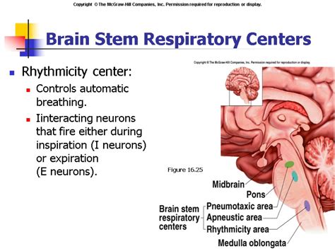
Table of Contents
Where is the Breathing Center Located? A Deep Dive into Respiratory Control
The simple act of breathing, something we take for granted thousands of times a day, is actually a complex process orchestrated by a sophisticated network within our brainstem. This network, often referred to as the respiratory center, isn't a single, clearly defined area but rather a collection of interconnected nuclei that work together to regulate our breathing patterns. Understanding its location and functionality is crucial for comprehending various respiratory disorders and their treatments.
The Brainstem: The Home of Respiratory Control
The primary location of the respiratory center is in the brainstem, specifically within the medulla oblongata and the pons. These are two vital parts of the brainstem, located at the base of the brain, connecting it to the spinal cord.
Medulla Oblongata: The Rhythm Generator
The medulla oblongata houses two crucial respiratory centers:
-
Dorsal Respiratory Group (DRG): This group is considered the primary rhythm generator for breathing. It's located in the dorsal part of the medulla and primarily controls inspiration, the inhalation phase of breathing. Neurons within the DRG fire rhythmically, sending signals to the phrenic nerves (which innervate the diaphragm) and intercostal nerves (which innervate the intercostal muscles). This rhythmic firing causes the diaphragm and intercostal muscles to contract, leading to lung expansion and air intake. The DRG receives sensory input from various receptors, allowing it to adjust breathing patterns based on body needs.
-
Ventral Respiratory Group (VRG): Situated in the ventral part of the medulla, the VRG is primarily active during forced breathing, such as during exercise or when you're struggling to catch your breath. It contains both inspiratory and expiratory neurons. While the DRG sets the basic rhythm, the VRG helps modify and fine-tune the breathing pattern, increasing the depth and rate of both inspiration and expiration when needed. It also plays a crucial role in coordinating the activity of the respiratory muscles.
Pons: Fine-Tuning Breathing
The pons, located superior to the medulla, contains two important respiratory centers that further refine the breathing process:
-
Pneumotaxic Center: This center acts as a "brake" on inspiration. It sends inhibitory signals to the DRG, limiting the duration of inspiration and thus controlling the rate and depth of breathing. By modulating the activity of the DRG, the pneumotaxic center helps prevent overinflation of the lungs. This ensures a smooth and controlled respiratory rhythm. A greater pneumotaxic center influence leads to shorter, more frequent breaths, whereas reduced activity results in slower, deeper breaths.
-
Apneustic Center: This center promotes inspiration. It sends stimulatory signals to the DRG, prolonging the inspiratory phase. However, its influence is modulated by the pneumotaxic center. The apneustic center's role is less clearly understood than the pneumotaxic center, but it's believed to contribute to the overall regulation of breathing depth and rate, particularly during periods of increased respiratory demand. An imbalance between the pneumotaxic and apneustic centers can lead to abnormal breathing patterns.
Sensory Input: Shaping the Respiratory Response
The respiratory center doesn't operate in isolation. It receives continuous feedback from various sensory receptors throughout the body:
-
Chemoreceptors: These receptors monitor the levels of oxygen (O2), carbon dioxide (CO2), and pH (acidity) in the blood. Changes in these parameters trigger appropriate adjustments in breathing rate and depth to maintain homeostasis. Peripheral chemoreceptors located in the carotid and aortic bodies are particularly sensitive to low O2 and high CO2 levels. Central chemoreceptors located in the medulla itself are sensitive to changes in CO2 and pH in the cerebrospinal fluid.
-
Mechanoreceptors: These receptors are located in the lungs and airways and respond to stretch and pressure. They provide information about lung volume and inflation. The Hering-Breuer reflex, mediated by these mechanoreceptors, helps prevent overinflation of the lungs by inhibiting further inspiration when the lungs are sufficiently expanded.
-
Proprioceptors: These receptors are located in the muscles and joints and provide information about body movement and position. During exercise, proprioceptive input stimulates increased breathing rate and depth to meet the increased oxygen demand.
Neural Pathways: Communication and Coordination
The respiratory center doesn't directly innervate all respiratory muscles. Instead, it relies on a complex network of neural pathways to communicate with these muscles:
-
Phrenic Nerve: This nerve originates in the cervical spinal cord and innervates the diaphragm, the primary muscle responsible for inspiration. Signals from the DRG and VRG travel down the spinal cord and through the phrenic nerve to stimulate diaphragm contraction.
-
Intercostal Nerves: These nerves originate in the thoracic spinal cord and innervate the intercostal muscles, which assist in both inspiration and expiration. The intercostal nerves receive signals from the DRG and VRG, coordinating their activity with that of the diaphragm.
-
Other Respiratory Muscles: The respiratory center also coordinates the activity of other accessory respiratory muscles, such as the sternocleidomastoid and scalene muscles, which become active during increased respiratory effort.
Clinical Significance: Understanding Respiratory Disorders
Understanding the location and function of the respiratory center is crucial for diagnosing and treating various respiratory disorders. Damage to the brainstem, for instance, can severely disrupt breathing patterns, leading to conditions like central apnea. Similarly, dysfunction of the chemoreceptors or other sensory input pathways can contribute to respiratory problems such as hypoventilation (decreased breathing) or hyperventilation (increased breathing).
Many neurological conditions, including stroke, trauma, and infections, can directly or indirectly affect the respiratory center, altering breathing patterns and potentially leading to life-threatening respiratory distress.
Conclusion: A Complex System Maintaining Life
The respiratory center, located primarily in the medulla and pons of the brainstem, is a remarkably complex system that orchestrates the essential process of breathing. Its intricate network of neurons, sensory inputs, and neural pathways work in concert to maintain an appropriate breathing pattern, adapting to the body's changing demands. Understanding its location and function is fundamental to comprehending the physiology of breathing and its relevance in clinical settings. The intricate interplay between the DRG, VRG, pneumotaxic, and apneustic centers, coupled with the continuous feedback from various receptors, ensures the seamless and life-sustaining regulation of our respiration. Further research continues to unravel the complexities of this vital control center and refine our understanding of respiratory function and dysfunction.
Latest Posts
Latest Posts
-
How Tall Is 62 Inches In Feet
Mar 15, 2025
-
Which Of The Following Are Correctly Matched
Mar 15, 2025
-
Whats The Square Root Of X
Mar 15, 2025
-
Whats The Square Root Of 128
Mar 15, 2025
-
72 In Is How Many Feet
Mar 15, 2025
Related Post
Thank you for visiting our website which covers about Where Is The Breathing Center Located . We hope the information provided has been useful to you. Feel free to contact us if you have any questions or need further assistance. See you next time and don't miss to bookmark.
