What Is The Difference Between Light And Electron Microscopes
Juapaving
Mar 17, 2025 · 6 min read
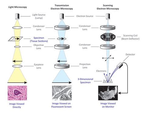
Table of Contents
What's the Difference Between Light and Electron Microscopes? A Deep Dive
Microscopes are indispensable tools in various scientific fields, allowing us to visualize the intricate details of the world invisible to the naked eye. However, not all microscopes are created equal. Two prominent types dominate the field: light microscopes and electron microscopes. While both aim to magnify images, their underlying principles, capabilities, and applications differ significantly. This comprehensive guide delves into the core distinctions between these powerful instruments, illuminating their unique strengths and limitations.
The Fundamentals: How Light and Electron Microscopes Work
The core difference between light and electron microscopes lies in the type of "light" they use to illuminate the specimen.
Light Microscopes: Harnessing Visible Light
Light microscopes, as their name suggests, utilize visible light to create magnified images. A light source illuminates the sample, and the light passing through (or reflected from) the specimen is then collected and magnified by a system of lenses. These lenses bend the light rays, creating a larger, virtual image that is perceived by the observer through the eyepiece or projected onto a screen. The maximum magnification achievable with a light microscope is typically around 1500x, limited by the wavelength of visible light.
Electron Microscopes: Exploring the Quantum Realm
Electron microscopes, on the other hand, use a beam of electrons instead of light to illuminate the specimen. Electrons, having a much shorter wavelength than visible light, allow for significantly higher resolution and magnification. The electron beam interacts with the sample, generating signals that are then processed to create an image. Because electrons are easily scattered by air molecules, electron microscopy necessitates a high-vacuum environment. Magnification in electron microscopes can reach several million times, revealing intricate ultrastructural details invisible to light microscopy.
Key Differences: A Comparative Analysis
The differences between light and electron microscopes extend beyond their fundamental illumination sources. Let's explore these crucial distinctions in more detail:
1. Resolution and Magnification: A Tale of Two Scales
Resolution, the ability to distinguish between two closely spaced objects, is a critical parameter in microscopy. Electron microscopes boast significantly superior resolution compared to light microscopes. This is because the wavelength of electrons is far shorter than that of visible light. Consequently, electron microscopes can resolve much finer details, revealing subcellular structures and even individual molecules.
Magnification refers to the enlargement of the image. While light microscopes can achieve magnifications up to 1500x, electron microscopes can magnify images millions of times, allowing for incredibly detailed observations of microscopic structures.
2. Sample Preparation: A Matter of Preservation
Sample preparation techniques differ substantially between the two microscope types.
Light Microscopy: Sample preparation for light microscopy is relatively straightforward. Many samples can be observed directly, or with minimal preparation, such as mounting on a slide. However, more complex techniques like staining or sectioning may be required to enhance contrast and visualize specific structures. Staining involves using dyes to selectively color different cellular components. Sectioning involves cutting very thin slices of the sample to allow light to pass through.
Electron Microscopy: Electron microscopy demands considerably more intricate sample preparation. Because the electron beam is easily scattered by air and water, samples must be meticulously dehydrated and embedded in resin. Thin sections are then cut using an ultramicrotome, producing incredibly thin slices (often just nanometers thick) that can be viewed under the electron beam. This process often involves chemical fixation, dehydration, embedding, sectioning, and staining with heavy metals to enhance contrast.
3. Imaging Modes: Diverse Approaches to Visualization
Both light and electron microscopes offer a variety of imaging modes, each providing unique insights into the sample's structure and composition.
Light Microscopy: Offers several modes like brightfield, darkfield, phase-contrast, and fluorescence microscopy. Brightfield microscopy is the most common, using transmitted light to illuminate the sample. Darkfield microscopy enhances contrast by illuminating the sample from the side, making transparent structures visible. Phase-contrast microscopy enhances contrast in transparent specimens by exploiting differences in refractive index. Fluorescence microscopy utilizes fluorescent dyes or proteins to label specific cellular components, enabling their visualization.
Electron Microscopy: Provides several specialized imaging techniques including Transmission Electron Microscopy (TEM) and Scanning Electron Microscopy (SEM). TEM transmits electrons through the sample, providing high-resolution images of internal structures. SEM scans the surface of the sample with a focused electron beam, generating high-resolution images of surface topography and three-dimensional structure. Other techniques like cryo-electron microscopy (cryo-EM) allow for imaging of samples in a near-native state, minimizing artifacts associated with conventional sample preparation.
4. Cost and Complexity: A Significant Difference
Electron microscopes are significantly more expensive and complex than light microscopes. Their sophisticated engineering, specialized equipment, and demanding sample preparation procedures contribute to their high cost. Furthermore, they require specialized training and expertise to operate effectively. Light microscopes, in contrast, are relatively inexpensive and user-friendly, making them accessible to a wider range of users and applications.
5. Applications: Tailored to Specific Needs
The choice between light and electron microscopy is heavily influenced by the nature of the research question and the desired level of detail.
Light Microscopy: Widely used in various fields including biology, medicine, and materials science. It's valuable for observing live cells, visualizing cellular processes, and studying the structure of tissues and organs at a relatively large scale. Its ease of use and versatility make it an ideal tool for educational purposes and routine analyses.
Electron Microscopy: Essential for high-resolution imaging in nanotechnology, materials science, and cell biology. Its ability to resolve subcellular structures and individual molecules makes it indispensable for studies of viruses, proteins, and other nanoscale entities. It's crucial for characterizing the structure and properties of materials at an atomic level.
Summary Table: A Quick Comparison
| Feature | Light Microscope | Electron Microscope |
|---|---|---|
| Illumination | Visible light | Electron beam |
| Resolution | Lower (limited by wavelength of light) | Much higher (limited by wavelength of electrons) |
| Magnification | Up to 1500x | Millions of times |
| Sample Prep | Relatively simple | Complex and time-consuming |
| Cost | Lower | Much higher |
| Complexity | Lower | Much higher |
| Applications | Biology, medicine, materials science (general) | Nanotechnology, materials science (high resolution), cell biology (ultrastructure) |
Choosing the Right Microscope: A Practical Guide
The optimal choice between a light and an electron microscope depends entirely on the specific research question and the required level of detail. If you need to observe live cells or study relatively large structures, a light microscope might suffice. However, if you need to visualize subcellular structures or individual molecules, an electron microscope is essential. Factors like budget, available expertise, and the complexity of sample preparation should also be considered when making this crucial decision. In some cases, a combination of both techniques may provide the most comprehensive understanding of the sample.
The Future of Microscopy: Innovation and Advancement
Microscopy continues to evolve, with ongoing advancements pushing the boundaries of resolution and imaging capabilities. Super-resolution light microscopy techniques, such as PALM and STORM, are bridging the gap between light and electron microscopy, achieving resolutions beyond the diffraction limit of light. Cryo-electron microscopy is revolutionizing structural biology, providing unprecedented insights into the structure and dynamics of macromolecular complexes. These ongoing developments promise even greater detail and understanding of the microscopic world in the years to come.
Latest Posts
Latest Posts
-
What Are The Characteristics Of Igneous Rocks
Mar 18, 2025
-
Circle How Many Lines Of Symmetry
Mar 18, 2025
-
What Is The Percentage Of 3 16
Mar 18, 2025
-
How Many Kings Are In A 52 Card Deck
Mar 18, 2025
-
What Is 3 8 In Percentage
Mar 18, 2025
Related Post
Thank you for visiting our website which covers about What Is The Difference Between Light And Electron Microscopes . We hope the information provided has been useful to you. Feel free to contact us if you have any questions or need further assistance. See you next time and don't miss to bookmark.
