What Is The Difference Between A Light And Electron Microscope
Juapaving
Apr 07, 2025 · 6 min read
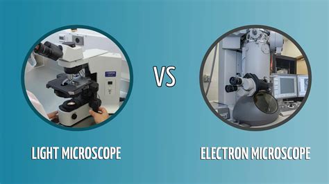
Table of Contents
Delving Deep: What Sets Light and Electron Microscopes Apart
Microscopes, the quintessential tools of scientific exploration, have revolutionized our understanding of the microscopic world. From the intricate structures of plant cells to the subatomic landscapes of materials, microscopes have enabled us to visualize worlds invisible to the naked eye. However, not all microscopes are created equal. This article delves into the fundamental differences between two dominant types: light microscopes and electron microscopes, exploring their principles, capabilities, and limitations. Understanding these distinctions is crucial for selecting the appropriate instrument for a given research application.
The Fundamentals of Light Microscopy
Light microscopy, the more established technique, utilizes visible light to illuminate the specimen and create a magnified image. Its simplicity and relative affordability have contributed to its widespread use in various fields, including biology, medicine, and materials science.
How Light Microscopes Work: A Journey Through Lenses
The basic principle hinges on refraction, the bending of light as it passes through different media (like air and glass). A typical light microscope employs a series of lenses—the objective lens positioned near the specimen, and the eyepiece lens (or ocular lens) through which the observer views the magnified image. The objective lens produces a real, inverted, and magnified image of the specimen, which is then further magnified by the eyepiece lens to produce a virtual image.
Types of Light Microscopy: Beyond the Basic
While the fundamental principle remains consistent, various techniques enhance the capabilities of light microscopy:
-
Bright-field microscopy: This is the most common type, where light passes directly through the specimen. Staining techniques are often employed to enhance contrast, as many biological samples are naturally transparent.
-
Dark-field microscopy: In this technique, only scattered light reaches the objective lens, creating a bright specimen against a dark background. This is particularly useful for observing unstained, transparent specimens.
-
Phase-contrast microscopy: This advanced method enhances the contrast of transparent specimens by exploiting differences in refractive index. This allows visualization of internal structures without the need for staining.
-
Fluorescence microscopy: This powerful technique utilizes fluorescent dyes or proteins that emit light at specific wavelengths when excited by a light source. It is widely used in biological research to visualize specific cellular structures or molecules.
-
Confocal microscopy: A sophisticated variant of fluorescence microscopy, confocal microscopy uses a pinhole aperture to eliminate out-of-focus light, resulting in highly detailed three-dimensional images.
Magnification and Resolution: The Limits of Light
A crucial aspect of any microscope is its magnification, representing the increase in the apparent size of the specimen. However, magnification alone is insufficient. Resolution, the ability to distinguish between two closely spaced points, is equally critical. The resolution of a light microscope is limited by the diffraction of light, which prevents the precise visualization of structures smaller than roughly half the wavelength of visible light (around 200 nanometers). This limitation restricts the detail achievable with light microscopy.
Electron Microscopy: Unveiling the Ultrastructure
Electron microscopy represents a significant leap in resolution and magnification. Instead of visible light, it utilizes a beam of electrons to illuminate the specimen. Because electrons have a much shorter wavelength than light, electron microscopes achieve far superior resolution, allowing visualization of structures at the nanometer scale.
Electron Microscopy Principles: A Quantum Leap
The fundamental principle of electron microscopy is the interaction of an electron beam with the specimen. Different types of electron microscopy exploit different aspects of this interaction:
-
Transmission Electron Microscopy (TEM): In TEM, a highly focused electron beam passes through an ultrathin specimen. The interaction of electrons with the specimen results in a pattern of transmitted and scattered electrons, which are then captured to create an image. TEM offers exceptionally high resolution, capable of resolving individual atoms. However, sample preparation for TEM is complex and time-consuming, requiring ultra-thin sections.
-
Scanning Electron Microscopy (SEM): SEM uses a focused electron beam that scans across the surface of the specimen. The interaction of the beam with the specimen generates various signals, including secondary electrons, backscattered electrons, and X-rays. These signals are used to create a three-dimensional image of the specimen's surface. SEM is excellent for visualizing surface features and topography, offering exceptional depth of field.
Magnification and Resolution: A Superior View
Electron microscopes achieve significantly higher magnification and resolution than light microscopes. TEM, in particular, can achieve resolutions down to sub-angstrom levels, revealing the detailed atomic structure of materials. SEM, while not as high-resolution as TEM, still offers superior resolution compared to light microscopy, enabling the visualization of fine surface details.
Sample Preparation: The Crucial Step
Sample preparation is a critical aspect of electron microscopy, often requiring specialized techniques. For TEM, the specimen must be extremely thin (often less than 100 nanometers) to allow the electron beam to penetrate. This often involves embedding the specimen in resin, sectioning it using an ultramicrotome, and staining it with heavy metals to enhance contrast. SEM sample preparation is generally less demanding but may still require coating the specimen with a conductive material to prevent charging.
A Comparative Overview: Light vs. Electron Microscopy
The table below summarizes the key differences between light and electron microscopes:
| Feature | Light Microscope | Electron Microscope |
|---|---|---|
| Illumination | Visible light | Electron beam |
| Wavelength | 400-700 nm | <0.004 nm (TEM) |
| Resolution | ~200 nm | <0.1 nm (TEM), ~1 nm (SEM) |
| Magnification | Up to 1500x | Up to 1,000,000x (TEM) |
| Sample Prep. | Relatively simple | Complex, often requires specialized techniques |
| Cost | Relatively inexpensive | Very expensive |
| Sample | Can be live or fixed | Usually fixed, dehydrated, and often coated |
| Image Type | 2D (mostly), some 3D techniques available | 2D or 3D, depending on the type |
| Applications | Biology, medicine, materials science (general) | Materials science, nanotechnology, cell biology (ultrastructure) |
Conclusion: Choosing the Right Tool
The choice between light and electron microscopy depends entirely on the research question and the nature of the specimen. Light microscopy offers simplicity, affordability, and the ability to observe live specimens. However, its resolution is limited. Electron microscopy, with its significantly higher resolution, is indispensable for visualizing ultrastructural details at the nanometer and sub-nanometer scales. While more complex and expensive, electron microscopy unlocks a world of detail invisible to light microscopy, pushing the boundaries of scientific discovery. Often, researchers use a combination of both techniques to gain a comprehensive understanding of the specimen under investigation. The power of microscopy lies not only in the technology itself but also in the skilled hands and insightful minds that wield these powerful tools.
Latest Posts
Latest Posts
-
What Is The Difference Between Cell Wall And Cell Membrane
Apr 08, 2025
-
How Many Hours In A Leap Year
Apr 08, 2025
-
What Is The Lcm Of 6 12 And 15
Apr 08, 2025
-
47 Inches Is How Many Feet
Apr 08, 2025
-
Can A Negative Number Be Rational
Apr 08, 2025
Related Post
Thank you for visiting our website which covers about What Is The Difference Between A Light And Electron Microscope . We hope the information provided has been useful to you. Feel free to contact us if you have any questions or need further assistance. See you next time and don't miss to bookmark.