What Colour Is An Animal Cell
Juapaving
Mar 20, 2025 · 5 min read
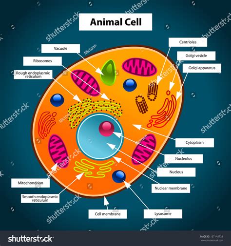
Table of Contents
What Color Is an Animal Cell? The Surprising Answer and the Science Behind It
The question, "What color is an animal cell?" might seem deceptively simple. After all, we've all seen colorful diagrams in textbooks, right? However, the reality is far more nuanced and fascinating. The short answer is: animal cells don't have a specific color. Their appearance depends on a multitude of factors, and understanding this requires a deeper dive into cell biology and microscopy.
The Illusion of Color in Textbook Diagrams
The vibrant colors you see in biology textbooks are purely artistic representations. They're used to highlight different cellular structures and make them easier to understand. These diagrams aren't photographic representations of actual cells. In reality, observing unstained animal cells under a light microscope reveals a largely colorless or translucent appearance.
This lack of inherent color is because the main components of an animal cell – the cytoplasm, nucleus, mitochondria, and other organelles – are largely composed of water, proteins, and lipids. These substances don't absorb or reflect visible light in a way that creates a distinct hue.
Microscopic Techniques and the Appearance of Color
To visualize animal cells and their internal structures, scientists employ various microscopic techniques that introduce color artificially. These techniques are crucial for understanding cell biology and are not reflective of the cell's natural color.
1. Staining Techniques
The most common approach is using stains, which are dyes that bind to specific cellular components. Different stains target different molecules, allowing researchers to differentiate various organelles and structures.
-
Hematoxylin and Eosin (H&E) staining: This is a widely used staining technique in histology (the study of tissues). Hematoxylin stains nuclei a deep purple or blue, while eosin stains the cytoplasm and extracellular matrix pink or red. This gives a clear contrast and allows for identification of different cell types and their structures.
-
Specific stains for organelles: Other stains target specific organelles like mitochondria (using dyes like Janus Green), Golgi apparatus, or the endoplasmic reticulum. These specialized stains provide a more detailed view of the cell's internal machinery, but again, the colors are artificial.
The colors observed after staining are determined by the chemical properties of the stain and its interaction with the cellular components, not the inherent color of the cell itself.
2. Fluorescent Microscopy
Fluorescent microscopy uses fluorescent dyes or proteins (like GFP – Green Fluorescent Protein) that emit light at specific wavelengths when excited by a light source. This technique allows visualization of specific molecules or structures within a cell with high sensitivity.
While this technique provides stunning images, often showcasing vibrant greens, reds, or other colors, these colors are due to the fluorescent molecules used, not the inherent color of the animal cell.
3. Electron Microscopy
Electron microscopy uses a beam of electrons instead of light to create images. This technique provides incredibly high resolution, revealing cellular structures with much greater detail than light microscopy. However, electron microscopy doesn't inherently show color. The resulting images are grayscale, and color is often added artificially later for interpretation.
Factors Influencing the Apparent Color of Animal Cell Aggregates (Tissues and Organs)
While individual animal cells are largely colorless, the apparent color of tissues and organs is influenced by various factors. This apparent color isn't due to the cells themselves, but rather the substances within and around them.
-
Blood: The presence of blood, rich in hemoglobin (a red pigment carrying oxygen), contributes significantly to the color of many tissues and organs. Muscles, for instance, appear red due to their rich blood supply.
-
Pigments: Some cells contain pigments that contribute to the color of tissues. Melanin, a brown-black pigment responsible for skin and hair color, is produced by specialized cells called melanocytes. Other pigments like carotenoids (yellow-orange) and anthocyanins (red-purple) can also influence tissue color. These pigments are contained within specific organelles or vesicles and don't represent the inherent color of the cell itself.
-
Lipofuscin: This yellowish-brown pigment accumulates in cells with age, and it contributes to the coloration of tissues in older individuals.
-
Bile pigments: These pigments, derived from the breakdown of hemoglobin, give a yellowish tint to some tissues, including the skin in cases of jaundice.
Therefore, the color of tissues and organs is a complex interplay of blood content, pigments produced by specialized cells, accumulated waste products, and other factors, not a reflection of the inherent color of the animal cells that compose them.
The Importance of Understanding Cell Color (or Lack Thereof)
Understanding that animal cells don't possess inherent color is crucial for several reasons:
-
Accurate interpretation of microscopic images: Researchers must be aware that the colors observed in stained or fluorescently labeled cells are artificial and reflect the dyes or labels used, not the cells' natural state.
-
Avoidance of misinterpretations: Confusing the artificially introduced colors in microscopy with the actual color of the cell can lead to erroneous conclusions.
-
Development of improved imaging techniques: Knowing the limitations of current techniques helps drive research into developing new methods to visualize cellular structures more accurately and with better resolution.
-
Advancement of cell biology: A fundamental understanding of cell composition and structure is essential for advancements in various areas of biological research, including disease diagnosis, drug development, and tissue engineering.
Conclusion: Beyond the Simple Question
The seemingly simple question of an animal cell's color leads to a fascinating exploration of cell biology, microscopy techniques, and the diverse factors influencing tissue coloration. While individual cells appear colorless or translucent under a light microscope, the vibrant hues seen in diagrams and microscopy images are the result of artificial staining and labeling methods. Understanding this distinction is vital for accurate interpretation and advancements in the field of cell biology. The true beauty of an animal cell lies not in its color, but in its intricate structure and function, a world revealed only through the application of advanced scientific techniques.
Latest Posts
Latest Posts
-
5 Out Of 8 As A Percentage
Mar 21, 2025
-
Boiling Point Of Water Kelvin Scale
Mar 21, 2025
-
150 Cm Is How Many Inches
Mar 21, 2025
-
120 Sq Mt To Sq Ft
Mar 21, 2025
-
What Is 19 25 As A Percent
Mar 21, 2025
Related Post
Thank you for visiting our website which covers about What Colour Is An Animal Cell . We hope the information provided has been useful to you. Feel free to contact us if you have any questions or need further assistance. See you next time and don't miss to bookmark.
