Mitosis Of An Onion Root Tip
Juapaving
Mar 12, 2025 · 7 min read
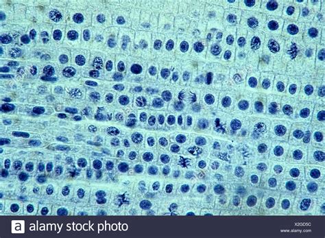
Table of Contents
Mitosis in Onion Root Tips: A Comprehensive Guide
The humble onion, Allium cepa, is more than just a culinary staple; it's a surprisingly valuable tool for understanding fundamental biological processes. Specifically, the rapidly dividing cells found in the root tip of an onion provide an excellent model system for studying mitosis, the process of cell division that's crucial for growth and repair in all eukaryotic organisms. This detailed guide will explore the intricacies of observing and understanding mitosis in onion root tips, covering everything from preparation techniques to identifying the different phases.
Why Onion Root Tips?
Onion root tips are ideal for observing mitosis for several key reasons:
-
High mitotic index: The root tip contains a meristematic region, a zone of actively dividing cells. This high mitotic index ensures a readily observable number of cells undergoing mitosis at any given time. This significantly simplifies the process of finding cells in different stages.
-
Ease of access and preparation: Onion roots are readily available, inexpensive, and relatively easy to prepare for microscopic observation. The procedure doesn't require extensive laboratory equipment or specialized skills, making it suitable for educational settings.
-
Clear chromosomal structure: The chromosomes in onion root tip cells are relatively large and well-defined, making them easily visible under a light microscope. This clarity is crucial for accurate identification of the different phases of mitosis.
-
Ethical considerations: Using onion root tips eliminates any ethical concerns associated with using animal tissues or cells. This makes it a preferred choice for many educational and research purposes.
Preparing Onion Root Tips for Microscopic Observation
Before we delve into the stages of mitosis, let's outline the steps involved in preparing the onion root tips for microscopic examination. The key is to halt the cells in the various stages of mitosis to allow for clear visualization.
1. Growing the Roots
To obtain suitable root tips, you'll need to grow onions. Simply place the bottom of an onion (the part with the roots) in a shallow dish of water. Ensure only the base of the onion is submerged; otherwise, the onion might rot. After a few days (typically 3-5), roots approximately 2-3 cm long will develop. These young, actively growing roots are rich in actively dividing cells.
2. Pretreatment (Hydrolysis)
This crucial step stops the cell cycle in its tracks, preventing further division and spreading the chromosomes more easily for observation. A common pretreatment involves using a chemical fixative and then hydrolyzing the roots in a diluted acid. Hydrolysis aids in the loosening of the tissues, so chromosomes separate more easily.
Important Note: The exact protocols for pretreatment vary, but generally involve solutions such as hydrochloric acid (HCl) at specific concentrations and for specific durations. Always prioritize safety and handle chemicals with appropriate precautions, using gloves and eye protection.
3. Fixing
Fixing preserves the cellular structures, preventing further cellular changes and degradation. This step uses a fixative, typically a combination of alcohol and acetic acid, which crosslinks proteins and stabilizes cellular components. Again, exact protocols should be followed carefully to prevent damage to the samples.
4. Staining
After fixing and washing, the root tips are stained to improve the visibility of the chromosomes. Common stains include acetocarmine or Feulgen stain. These stains bind to DNA, making the chromosomes intensely colored and easily distinguishable under the microscope.
5. Squashing
To create a thin, easily observable sample, the stained root tips are carefully squashed between a slide and coverslip. This process spreads the cells thinly to observe individual chromosomes in detail. Gentle pressure is vital to avoid damaging the cells and smearing the chromosomes.
Stages of Mitosis in Onion Root Tips
Mitosis is a continuous process, but for descriptive purposes, it's divided into several distinct phases:
1. Prophase
- Characteristics: The chromatin condenses into visible, distinct chromosomes. Each chromosome consists of two identical sister chromatids joined at the centromere. The nuclear envelope begins to break down, and the mitotic spindle starts to form.
- Microscopic observation: You'll observe thickened, rod-shaped chromosomes scattered throughout the cell. The nucleolus, a dense structure within the nucleus, is usually no longer visible.
2. Prometaphase
- Characteristics: The nuclear envelope completely disintegrates. The chromosomes continue to condense and move towards the center of the cell. Kinetochores, protein structures at the centromeres, attach to the microtubules of the mitotic spindle.
- Microscopic observation: Chromosomes are clearly visible and appear to be moving towards the cell's equator.
3. Metaphase
- Characteristics: The chromosomes align along the metaphase plate, an imaginary plane equidistant from the two poles of the spindle. Each chromosome is attached to microtubules from both poles. This alignment ensures accurate segregation of chromosomes during the subsequent phases.
- Microscopic observation: Chromosomes are neatly arranged in a single line across the center of the cell. This is a key identifying characteristic of metaphase.
4. Anaphase
- Characteristics: The sister chromatids separate at the centromere, and each chromatid (now considered a chromosome) moves towards opposite poles of the cell. The microtubules shorten, pulling the chromosomes along.
- Microscopic observation: The chromosomes appear to be pulled apart, moving towards opposite ends of the cell. The V-shaped appearance of the chromosomes is characteristic of anaphase.
5. Telophase
- Characteristics: The chromosomes arrive at the poles of the cell. The chromosomes decondense, becoming less visible. The nuclear envelope reforms around each set of chromosomes, forming two new nuclei. The spindle apparatus disassembles.
- Microscopic observation: Two distinct nuclei are visible at opposite ends of the cell. Chromosomes are less defined than in previous phases.
6. Cytokinesis
- Characteristics: Cytokinesis is not technically part of mitosis but is the final step in the cell cycle. It's the division of the cytoplasm, resulting in two separate daughter cells, each with a complete set of chromosomes. In plant cells, a cell plate forms in the middle of the cell, eventually developing into a new cell wall.
- Microscopic observation: A cell plate (in plant cells) or a constriction (in animal cells) will be visible, indicating the division of the cytoplasm. Eventually, two distinct daughter cells will be seen.
Identifying Stages under the Microscope
Successfully identifying the different stages of mitosis requires practice and careful observation. Start by focusing on key features of each phase:
- Prophase: Chromosomes are condensing; nuclear envelope is breaking down.
- Metaphase: Chromosomes are aligned at the metaphase plate.
- Anaphase: Sister chromatids are separating.
- Telophase: Two distinct nuclei are forming.
- Cytokinesis: Cell division is complete.
Calculating the Mitotic Index
The mitotic index is the ratio of the number of cells undergoing mitosis to the total number of cells observed. It provides valuable insight into the rate of cell division in the tissue. To calculate it:
- Count the total number of cells in your microscopic field of view.
- Count the number of cells in each phase of mitosis.
- Calculate the mitotic index using the formula:
(Number of cells in mitosis / Total number of cells) x 100%
Beyond the Basics: Further Exploration
The study of mitosis in onion root tips serves as a fundamental introduction to cell biology. Further exploration can involve:
- Investigating the effects of environmental factors: Explore how temperature, light, or chemical treatments affect the mitotic index.
- Comparing mitosis in different plant species: Observe mitosis in other plant root tips and compare the characteristics.
- Exploring the role of specific proteins: Investigate the functions of key proteins involved in the regulation of mitosis.
Conclusion
Observing mitosis in onion root tips is a classic and accessible experiment that provides invaluable insight into this fundamental biological process. By following the preparation techniques outlined above and carefully observing the characteristic features of each stage, you can gain a deeper understanding of how cells divide, ensuring growth, repair, and the continuity of life. Remember safety precautions when handling chemicals, and enjoy the fascinating world of cellular division. The seemingly simple onion holds a wealth of biological secrets just waiting to be uncovered.
Latest Posts
Latest Posts
-
If X Is A Multiple Of 18 And 60
May 09, 2025
-
Does Light Require A Medium To Travel
May 09, 2025
-
Cuanto Es 15 Inches En Centimetros
May 09, 2025
-
The Least Common Multiple Of 12 And 8
May 09, 2025
-
Is 5 7 A Rational Number
May 09, 2025
Related Post
Thank you for visiting our website which covers about Mitosis Of An Onion Root Tip . We hope the information provided has been useful to you. Feel free to contact us if you have any questions or need further assistance. See you next time and don't miss to bookmark.