Label The Endocrine Glands On The Figure
Juapaving
Mar 28, 2025 · 7 min read
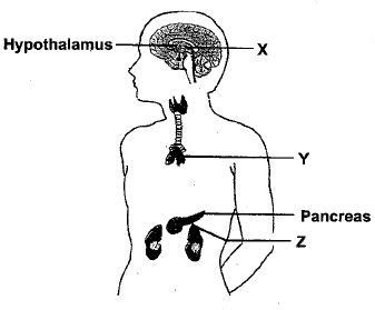
Table of Contents
Label the Endocrine Glands on the Figure: A Comprehensive Guide to the Human Endocrine System
The endocrine system, a complex network of glands and hormones, plays a vital role in regulating various bodily functions. Understanding its components is crucial for comprehending human physiology. This article serves as a comprehensive guide to identifying and understanding the major endocrine glands, providing detailed information to effectively "label the endocrine glands on the figure" – a common task in anatomy and physiology studies. We will explore each gland's location, function, and the hormones it produces, equipping you with a thorough understanding of this essential system.
The Major Endocrine Glands: Location and Function
Before we dive into labeling, let's familiarize ourselves with the key players of the endocrine system. Remember, this is not an exhaustive list, but it covers the major glands typically included in introductory anatomy and physiology diagrams.
1. Hypothalamus: The Master Regulator
Located in the brain, the hypothalamus is the master control center of the endocrine system. While not technically a gland itself, it produces releasing and inhibiting hormones that regulate the pituitary gland. These hormones control the anterior pituitary's hormone secretion, influencing growth, metabolism, reproduction, and stress response. Think of it as the conductor of an orchestra, coordinating the activities of other endocrine glands.
Key Hormones Influenced by the Hypothalamus: Gonadotropin-releasing hormone (GnRH), thyrotropin-releasing hormone (TRH), corticotropin-releasing hormone (CRH), growth hormone-releasing hormone (GHRH), somatostatin, dopamine.
2. Pituitary Gland (Hypophysis): The "Master Gland"
Situated beneath the hypothalamus, the pituitary gland is often called the "master gland" because it controls many other endocrine glands. It has two distinct lobes: the anterior and posterior pituitary.
-
Anterior Pituitary: Produces several crucial hormones, including growth hormone (GH), prolactin (PRL), thyroid-stimulating hormone (TSH), adrenocorticotropic hormone (ACTH), follicle-stimulating hormone (FSH), and luteinizing hormone (LH). These hormones regulate growth, milk production, thyroid function, adrenal cortex function, and reproduction.
-
Posterior Pituitary: Stores and releases hormones produced by the hypothalamus – oxytocin and antidiuretic hormone (ADH or vasopressin). Oxytocin plays a role in childbirth and lactation, while ADH regulates water balance.
3. Thyroid Gland: Metabolism and Development
Located in the neck, the thyroid gland produces thyroid hormones – thyroxine (T4) and triiodothyronine (T3). These hormones are essential for regulating metabolism, growth, and development. They influence energy expenditure, heart rate, and body temperature. The thyroid gland also produces calcitonin, a hormone that helps regulate calcium levels in the blood.
4. Parathyroid Glands: Calcium Regulation
Four small parathyroid glands are embedded in the back of the thyroid gland. They secrete parathyroid hormone (PTH), which plays a vital role in maintaining calcium and phosphorus levels in the blood. PTH increases blood calcium levels by stimulating bone resorption, increasing calcium absorption in the intestines, and enhancing calcium reabsorption in the kidneys. It acts antagonistically to calcitonin.
5. Adrenal Glands: Stress Response and Metabolism
The adrenal glands, situated atop the kidneys, consist of two parts: the adrenal cortex and the adrenal medulla.
-
Adrenal Cortex: Produces steroid hormones, including glucocorticoids (cortisol), mineralocorticoids (aldosterone), and androgens. Cortisol regulates stress response, glucose metabolism, and inflammation. Aldosterone regulates sodium and potassium balance. Androgens contribute to secondary sexual characteristics.
-
Adrenal Medulla: Produces catecholamines, primarily epinephrine (adrenaline) and norepinephrine (noradrenaline). These hormones are involved in the "fight-or-flight" response, increasing heart rate, blood pressure, and glucose levels in response to stress.
6. Pineal Gland: Circadian Rhythms
Located in the brain, the pineal gland produces melatonin, a hormone that regulates sleep-wake cycles (circadian rhythms). Melatonin secretion is influenced by light exposure, with increased production occurring in darkness.
7. Pancreas: Blood Sugar Regulation
The pancreas is both an exocrine and endocrine gland. Its endocrine function involves the production of insulin and glucagon by the islets of Langerhans. Insulin lowers blood glucose levels, while glucagon raises them. This delicate balance is crucial for maintaining blood sugar homeostasis.
8. Ovaries (Females): Reproduction and Secondary Sexual Characteristics
The ovaries, located in the pelvic cavity, produce estrogen and progesterone. Estrogen is crucial for the development of female secondary sexual characteristics and the regulation of the menstrual cycle. Progesterone prepares the uterus for pregnancy.
9. Testes (Males): Reproduction and Secondary Sexual Characteristics
The testes, located in the scrotum, produce testosterone, the primary male sex hormone. Testosterone is responsible for the development of male secondary sexual characteristics, sperm production, and the maintenance of male reproductive function.
Labeling the Endocrine Glands: A Practical Approach
Now that we've reviewed the major endocrine glands, let's discuss how to effectively label them on a diagram.
1. Understand the Diagram: Before you begin labeling, carefully examine the diagram. Identify the key anatomical structures surrounding the endocrine glands. This will help you correctly identify the location of each gland.
2. Use Clear and Concise Labels: Use short, unambiguous labels that clearly identify each gland. For example, use "Hypothalamus" instead of "Hypothalamic Region" or "Pituitary Gland" instead of "Anterior and Posterior Pituitary."
3. Accurate Placement of Labels: Position labels clearly and avoid overlapping other structures. Use leader lines (connecting lines) to connect each label to its corresponding gland. This ensures clarity and prevents ambiguity.
4. Consistent Formatting: Maintain consistent font size, style, and color for all labels to create a professional and readable figure.
5. Check for Completeness: Once you have labeled all glands, review your work to ensure that you have accurately identified and labeled all the structures indicated in the diagram.
Example Labeling (for a typical diagram):
- Hypothalamus: Label this structure in the brain, indicating its relationship to the pituitary gland.
- Anterior Pituitary: Clearly label the anterior lobe of the pituitary gland, distinguishing it from the posterior lobe.
- Posterior Pituitary: Label the posterior lobe, indicating its connection to the hypothalamus.
- Thyroid Gland: Label the butterfly-shaped gland located in the neck.
- Parathyroid Glands: Indicate the small glands embedded on the posterior surface of the thyroid.
- Adrenal Glands: Label the glands located atop the kidneys, perhaps with sub-labels for the cortex and medulla if the detail is present.
- Pancreas: Label the pancreas and if possible, its islets of Langerhans (if detailed enough).
- Ovaries (female diagram): Label the ovaries in the pelvic region.
- Testes (male diagram): Label the testes in the scrotum.
- Pineal Gland: Label this gland within the brain.
Beyond the Basics: Advanced Considerations
Understanding the interconnections between glands and the feedback loops that regulate hormone levels is crucial for a complete grasp of the endocrine system. For example, the hypothalamus regulates the pituitary, which in turn regulates other glands like the thyroid and adrenal glands. These interactions are often depicted in flowcharts or diagrams in more advanced studies.
Furthermore, understanding the clinical implications of endocrine disorders is also important. Conditions such as hypothyroidism, hyperthyroidism, diabetes mellitus, Cushing's syndrome, and Addison's disease all arise from dysfunctions within the endocrine system. Studying these conditions in relation to the glands involved helps build a deeper understanding.
Finally, the study of endocrine disruptors – environmental chemicals that interfere with hormone function – is a growing field. Understanding how these substances can affect the endocrine system's delicate balance highlights the importance of maintaining endocrine health.
Conclusion: Mastering the Endocrine System
Labeling the endocrine glands on a figure is a fundamental step in understanding the complexities of the human endocrine system. This process requires not only accurate identification of the glands but also a comprehensive understanding of their locations, functions, and the hormones they produce. This detailed guide provides a solid foundation for mastering this essential aspect of human anatomy and physiology. Remember to use clear, concise labels, accurately position them on the diagram, and ensure that the diagram is complete and easy to understand. By thoroughly understanding the endocrine system, you can build a strong foundation for further study in biology, medicine, and related fields. Furthermore, this knowledge fosters a deeper appreciation for the intricate mechanisms that govern our bodies and maintain overall health.
Latest Posts
Latest Posts
-
Which Of The Following Is Correctly Matched A
Mar 31, 2025
-
The Temperature At Which A Solid Becomes A Liquid
Mar 31, 2025
-
How Many Pairs Of Homologous Chromosomes Do Females Have
Mar 31, 2025
-
54 As Product Of Prime Factors
Mar 31, 2025
-
Organelles That Are The Sites Of Protein Synthesis
Mar 31, 2025
Related Post
Thank you for visiting our website which covers about Label The Endocrine Glands On The Figure . We hope the information provided has been useful to you. Feel free to contact us if you have any questions or need further assistance. See you next time and don't miss to bookmark.
