Function Of The Motor End Plate
Juapaving
Mar 17, 2025 · 6 min read
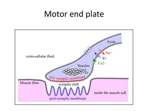
Table of Contents
The Motor End Plate: A Deep Dive into Neuromuscular Junction Function
The motor end plate, also known as the neuromuscular junction (NMJ), is a critical structure that facilitates communication between motor neurons and skeletal muscle fibers. This specialized synapse ensures the efficient and precise transmission of nerve impulses, leading to muscle contraction. Understanding its intricate function is crucial for comprehending movement, and various neurological and muscular disorders. This comprehensive article will explore the detailed anatomy, physiology, and clinical significance of the motor end plate.
Anatomy of the Neuromuscular Junction
The NMJ is a highly organized structure composed of several key components:
1. The Presynaptic Terminal:
This is the axon terminal of a motor neuron, the final segment of the nerve fiber responsible for transmitting the signal. It's characterized by a high concentration of synaptic vesicles. These vesicles are tiny, membrane-bound sacs that store and release the neurotransmitter acetylcholine (ACh). The presynaptic terminal also contains voltage-gated calcium channels (Ca2+ channels) critical for ACh release. The arrival of an action potential at the presynaptic terminal triggers the opening of these channels, allowing an influx of Ca2+ ions. This calcium influx is the crucial trigger for exocytosis – the process by which synaptic vesicles fuse with the presynaptic membrane and release ACh into the synaptic cleft.
2. The Synaptic Cleft:
This is the narrow space (approximately 20-30 nm) separating the presynaptic terminal from the postsynaptic membrane of the muscle fiber. It's filled with extracellular matrix and contains enzymes, such as acetylcholinesterase (AChE), which plays a vital role in terminating the signal by rapidly breaking down ACh. The short distance across the synaptic cleft ensures efficient diffusion of ACh to its receptors on the muscle fiber.
3. The Postsynaptic Membrane:
Also known as the motor end-plate membrane, this specialized region of the muscle fiber's sarcolemma (plasma membrane) is highly folded, forming deep junctional folds. This folding significantly increases the surface area for ACh receptors. The postsynaptic membrane is densely packed with nicotinic acetylcholine receptors (nAChRs), ligand-gated ion channels that bind ACh. These receptors are crucial for converting the chemical signal (ACh) back into an electrical signal in the muscle fiber. When ACh binds to these receptors, the channels open, allowing the influx of sodium ions (Na+) and efflux of potassium ions (K+), resulting in depolarization of the muscle fiber membrane. This depolarization, known as the end-plate potential (EPP), initiates an action potential that spreads along the muscle fiber, ultimately leading to muscle contraction.
Physiology of Neuromuscular Transmission: A Step-by-Step Process
The process of neuromuscular transmission involves a precise sequence of events:
-
Nerve Impulse Arrival: A nerve impulse (action potential) travels down the motor neuron axon and reaches the presynaptic terminal.
-
Calcium Influx: The arrival of the action potential opens voltage-gated Ca2+ channels in the presynaptic membrane. Ca2+ ions rush into the presynaptic terminal.
-
Acetylcholine Release: The increased intracellular Ca2+ concentration triggers the fusion of synaptic vesicles with the presynaptic membrane. This process, called exocytosis, releases ACh into the synaptic cleft.
-
Acetylcholine Binding: ACh diffuses across the synaptic cleft and binds to nAChRs on the postsynaptic membrane of the muscle fiber.
-
End-Plate Potential (EPP): nAChR activation opens the ion channels, causing an influx of Na+ ions and efflux of K+ ions. This creates a local depolarization of the muscle fiber membrane, known as the EPP. The EPP is a graded potential, meaning its amplitude is proportional to the amount of ACh released. A single action potential in the motor neuron typically releases enough ACh to generate an EPP that exceeds the threshold for action potential generation in the muscle fiber.
-
Muscle Fiber Action Potential: The EPP initiates an action potential in the muscle fiber membrane. This action potential propagates along the sarcolemma and into the T-tubules, triggering the release of calcium from the sarcoplasmic reticulum.
-
Muscle Contraction: The release of calcium from the sarcoplasmic reticulum initiates the sliding filament mechanism, leading to muscle fiber contraction.
-
Acetylcholine Degradation: AChE, located in the synaptic cleft, rapidly hydrolyzes ACh into choline and acetate. This terminates the signal and prevents continuous muscle contraction. Choline is then transported back into the presynaptic terminal to be resynthesized into ACh.
Clinical Significance of Motor End Plate Dysfunction
Disruptions in the function of the motor end plate can lead to various neuromuscular disorders. Some key examples include:
1. Myasthenia Gravis:
This autoimmune disease is characterized by the production of antibodies that target and destroy nAChRs at the NMJ. This leads to a decrease in the number of functional receptors, reducing the amplitude of the EPP and impairing neuromuscular transmission. Symptoms typically include muscle weakness and fatigue, particularly in muscles involved in eye movement, facial expression, and swallowing.
2. Lambert-Eaton Myasthenic Syndrome (LEMS):
LEMS is another autoimmune disorder affecting the NMJ, but in this case, antibodies target voltage-gated Ca2+ channels in the presynaptic terminal. This reduces the amount of ACh released, leading to a decrease in the EPP amplitude and muscle weakness. LEMS is often associated with small cell lung cancer.
3. Botulism:
Botulinum toxin, produced by Clostridium botulinum, is a potent neurotoxin that blocks ACh release at the NMJ. This prevents muscle contraction, resulting in paralysis. Botulinum toxin is used medically in small doses to treat certain muscle disorders, such as blepharospasm and cervical dystonia.
4. Curare Poisoning:
Curare is a plant-derived toxin that acts as a competitive antagonist of nAChRs. It binds to the receptors but doesn't activate them, thus blocking ACh binding and preventing neuromuscular transmission, resulting in paralysis.
Investigating Motor End Plate Function: Diagnostic Techniques
Several techniques are available to assess the function of the motor end plate and diagnose neuromuscular disorders:
1. Electromyography (EMG):
EMG measures the electrical activity of muscles. In neuromuscular disorders, EMG can reveal changes in muscle fiber action potentials, reflecting impaired neuromuscular transmission.
2. Nerve Conduction Studies (NCS):
NCS assesses the speed and efficiency of nerve impulse conduction. These studies can identify abnormalities in nerve function that may contribute to neuromuscular disorders.
3. Repetitive Nerve Stimulation (RNS):
RNS involves delivering repeated electrical stimuli to a nerve and observing the resulting muscle responses. In myasthenia gravis, RNS typically reveals a decrement in the amplitude of muscle responses with repetitive stimulation, indicating impaired neuromuscular transmission.
Conclusion: The Motor End Plate – A Critical Component of Movement
The motor end plate, a highly specialized synapse, is crucial for efficient and precise communication between motor neurons and skeletal muscle fibers. Its intricate structure and function are essential for normal movement. Understanding the detailed physiology of the NMJ is crucial for diagnosing and treating various neuromuscular disorders, which can significantly impact quality of life. Further research into the intricacies of this vital structure will continue to improve our understanding and treatment of these debilitating conditions. The study of the motor end plate continues to be a vibrant and important area of neuroscience research, promising future breakthroughs in both diagnostic and therapeutic strategies. The continued investigation of its components, its mechanisms of action, and its susceptibility to dysfunction offers potential for improved clinical outcomes in neurological and neuromuscular disease. Advanced imaging techniques, coupled with genetic analysis and proteomic studies, provide exciting avenues to explore the complex interplay of factors influencing neuromuscular junction function. This will undoubtedly lead to a more comprehensive understanding of health and disease, allowing for the development of targeted and efficacious therapies for the benefit of patients.
Latest Posts
Latest Posts
-
Are Lysosomes Only In Animal Cells
Mar 17, 2025
-
What Are The Prime Factors Of 47
Mar 17, 2025
-
Electronic Configuration Of Cr And Cu
Mar 17, 2025
-
What Is The Lowest Common Multiple Of 12 And 24
Mar 17, 2025
-
Lcm Of 9 12 And 15
Mar 17, 2025
Related Post
Thank you for visiting our website which covers about Function Of The Motor End Plate . We hope the information provided has been useful to you. Feel free to contact us if you have any questions or need further assistance. See you next time and don't miss to bookmark.
