Differences Between Cervical Thoracic And Lumbar Vertebrae
Juapaving
Apr 05, 2025 · 7 min read

Table of Contents
Delving Deep into the Differences: Cervical, Thoracic, and Lumbar Vertebrae
The human spine, a marvel of engineering, is composed of 33 vertebrae, divided into five distinct regions: cervical, thoracic, lumbar, sacral, and coccygeal. While the sacral and coccygeal vertebrae fuse together, the cervical, thoracic, and lumbar vertebrae retain their individual characteristics, each uniquely adapted to their specific functional roles. Understanding the anatomical differences between these three vertebral regions is crucial for comprehending spinal biomechanics, diagnosing spinal pathologies, and appreciating the overall complexity of the human musculoskeletal system. This comprehensive guide will delve into the detailed distinctions between cervical, thoracic, and lumbar vertebrae, highlighting their key structural features and functional implications.
Cervical Vertebrae: The Neck's Nimble Support
The cervical spine, comprised of seven vertebrae (C1-C7), supports the head and facilitates its remarkable range of motion. Its unique structure enables the delicate balance between mobility and stability required for head rotation, flexion, and extension.
Distinguishing Features of Cervical Vertebrae:
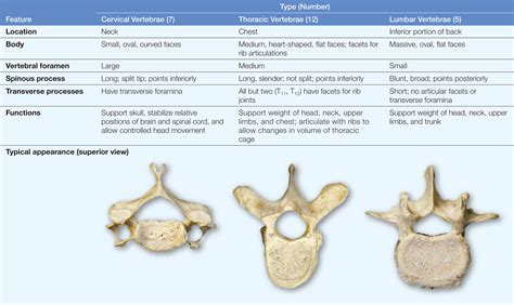
- Size and Shape: Cervical vertebrae are generally the smallest and most delicate of the three regions. Their bodies are relatively small and wider laterally than anteroposteriorly.
- Vertebral Foramen: The vertebral foramen (the opening through which the spinal cord passes) is relatively large in cervical vertebrae to accommodate the spinal cord's significant diameter in this region.
- Transverse Foramina: A defining characteristic of cervical vertebrae (except C7) are the presence of transverse foramina—holes in the transverse processes that allow the passage of the vertebral arteries and veins. These vessels supply blood to the brain.
- Spinous Processes: The spinous processes of cervical vertebrae are typically short and bifid (split into two), except for C7, which has a long, prominent spinous process easily palpable at the base of the neck. This is often referred to as the vertebra prominens.
- Articular Facets: The articular facets (surfaces where vertebrae articulate with each other) of the cervical vertebrae are oriented at an angle that allows for significant flexion, extension, and lateral bending. The superior articular facets face superiorly and slightly posteriorly, while the inferior articular facets face inferiorly and slightly anteriorly.
- Atlas (C1) and Axis (C2): The first two cervical vertebrae, the atlas (C1) and the axis (C2), are highly specialized. C1 lacks a body and has a ring-like structure that supports the skull, allowing for head rotation. C2 possesses the dens (odontoid process), a projection that fits into the atlas, enabling the pivoting motion of the head.
Thoracic Vertebrae: The Rib Cage's Firm Foundation
The twelve thoracic vertebrae (T1-T12) form the bony framework of the thorax, providing attachment points for the ribs and protecting vital organs such as the heart and lungs. This region prioritizes stability over mobility compared to the cervical spine.
Distinguishing Features of Thoracic Vertebrae:
- Size and Shape: Thoracic vertebrae are larger than cervical vertebrae but smaller than lumbar vertebrae. Their bodies are heart-shaped and gradually increase in size from T1 to T12.
- Vertebral Foramen: The vertebral foramen is relatively smaller than in the cervical spine, reflecting the reduced diameter of the spinal cord in this region.
- Costal Facets: Unique to thoracic vertebrae are the costal facets—articulating surfaces for the ribs. These facets are located on the vertebral bodies and transverse processes, allowing for the secure attachment of the ribs. The number and location of these facets vary depending on the individual vertebra. T1-T10 typically have two costal demi-facets on their vertebral bodies, articulating with the heads of two adjacent ribs. T11 and T12 possess a single, complete costal facet on their bodies.
- Spinous Processes: The spinous processes of thoracic vertebrae are long, pointed, and slope sharply downwards. This arrangement contributes to the thoracic spine's relative immobility.
- Articular Facets: The articular facets of thoracic vertebrae are oriented in a way that restricts excessive flexion and extension but allows for some rotation. The superior articular facets face posteriorly and laterally, while the inferior articular facets face anteriorly and medially.
Lumbar Vertebrae: The Lower Back's Weight-Bearing Powerhouse
The five lumbar vertebrae (L1-L5) are the largest and strongest of the vertebral column, bearing the weight of the upper body and transmitting it to the pelvis. They are adapted for weight-bearing and stability, prioritizing strength over mobility.
Distinguishing Features of Lumbar Vertebrae:
- Size and Shape: Lumbar vertebrae are the largest, with robust, kidney-shaped bodies. Their size reflects their substantial weight-bearing function.
- Vertebral Foramen: The vertebral foramen is relatively large compared to the thoracic vertebrae but smaller than the cervical vertebrae.
- Transverse Processes: The transverse processes of lumbar vertebrae are long and slender, lacking the transverse foramina found in cervical vertebrae.
- Spinous Processes: The spinous processes are thick, short, and relatively broad, projecting posteriorly.
- Articular Facets: The articular facets of lumbar vertebrae are oriented in a sagittal plane (vertical), facilitating flexion and extension but restricting rotation. The superior articular facets face medially, while the inferior articular facets face laterally.
- Mammillary Processes: Lumbar vertebrae possess mammillary processes, small projections located on the superior articular processes, which provide attachment points for muscles.
Comparative Table: Cervical, Thoracic, and Lumbar Vertebrae
To consolidate the differences, here’s a concise comparative table:
| Feature | Cervical Vertebrae | Thoracic Vertebrae | Lumbar Vertebrae |
|---|---|---|---|
| Size | Smallest | Medium | Largest |
| Body Shape | Wider laterally | Heart-shaped | Kidney-shaped |
| Vertebral Foramen | Largest | Medium | Large |
| Transverse Foramina | Present (except C7) | Absent | Absent |
| Spinous Process | Short, bifid (except C7) | Long, pointed, downward sloping | Short, thick, broad |
| Costal Facets | Absent | Present | Absent |
| Articular Facets Orientation | Allows flexion, extension, lateral bending | Restricts flexion & extension, allows rotation | Facilitates flexion & extension, restricts rotation |
| Special Features | Atlas (C1) & Axis (C2) | Costal facets | Mammillary processes |
Clinical Significance of Vertebral Differences
Understanding the distinct anatomical features of cervical, thoracic, and lumbar vertebrae is crucial in clinical settings. Different regions are susceptible to various pathologies, and recognizing the specific anatomical characteristics is essential for accurate diagnosis and treatment. For example:
- Cervical Spondylosis: Degenerative changes in the cervical spine, characterized by disc degeneration, osteophyte formation (bone spurs), and facet joint hypertrophy, often lead to neck pain, stiffness, and radiculopathy (nerve root compression).
- Thoracic Outlet Syndrome: Compression of nerves and blood vessels in the thoracic outlet (the space between the clavicle and first rib) can result in pain, numbness, and weakness in the arm and hand.
- Spinal Stenosis: Narrowing of the spinal canal, most commonly affecting the lumbar spine, can cause pain, numbness, weakness, and gait disturbances.
- Spondylolisthesis: Forward slippage of one vertebra over another, typically in the lumbar region, can lead to back pain, instability, and nerve compression.
- Scoliosis: Lateral curvature of the spine, affecting any region but often most prominent in the thoracic spine, can cause pain, deformity, and respiratory compromise.
Conclusion: A Symphony of Structure and Function
The human spine is a testament to the elegance and efficiency of biological design. The remarkable differences between cervical, thoracic, and lumbar vertebrae reflect their specific roles in supporting the body, protecting the spinal cord, and facilitating a wide range of movements. By understanding these differences, we gain a deeper appreciation for the intricate mechanics of the spine and the importance of maintaining its health and integrity. This knowledge is not only crucial for healthcare professionals but also for individuals seeking to understand their own bodies and make informed choices about their health and well-being. Further exploration into the complexities of the musculoskeletal system will only deepen our understanding of this extraordinary structure. Remember, maintaining good posture, engaging in regular exercise, and practicing proper lifting techniques are all essential steps in preserving the health of your spine for a lifetime.
Latest Posts
Latest Posts
-
What Is The Lateral Area Of A Rectangular Prism
Apr 06, 2025
-
Distinguish Between Centripetal Force And Centrifugal Force
Apr 06, 2025
-
What Is A Political Party Class 10
Apr 06, 2025
-
Which Of The Following Is An Even Function
Apr 06, 2025
-
How Do You Describe A Book
Apr 06, 2025
Related Post
Thank you for visiting our website which covers about Differences Between Cervical Thoracic And Lumbar Vertebrae . We hope the information provided has been useful to you. Feel free to contact us if you have any questions or need further assistance. See you next time and don't miss to bookmark.
