Diagram Of An Animal Cell And Plant Cell
Juapaving
Mar 17, 2025 · 5 min read
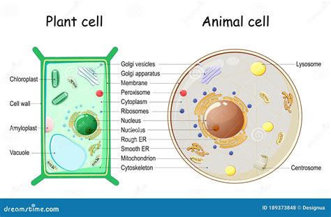
Table of Contents
Diagram of an Animal Cell and Plant Cell: A Comprehensive Comparison
Understanding the fundamental building blocks of life—cells—is crucial for anyone interested in biology. While all cells share some common features, significant differences exist between plant and animal cells. This detailed article will delve into the intricate structures of both, providing a comprehensive comparison illustrated with diagrams, highlighting their unique characteristics and functionalities.
The Animal Cell: A Dynamic Powerhouse
Animal cells, the basic units of animal tissues and organs, are eukaryotic cells, meaning they possess a membrane-bound nucleus and other organelles. Their structure is remarkably complex, supporting a wide range of functions essential for survival. Let's explore the key components:
1. The Cell Membrane (Plasma Membrane): The Gatekeeper
The cell membrane is the outer boundary of the animal cell, a selectively permeable barrier regulating the passage of substances in and out. This dynamic structure, composed primarily of a phospholipid bilayer with embedded proteins, controls the cell's internal environment, maintaining homeostasis. Proteins within the membrane act as channels, transporters, and receptors, facilitating communication and transport processes.
2. The Nucleus: The Control Center
The nucleus, the cell's largest and most prominent organelle, houses the cell's genetic material, DNA. DNA is organized into chromosomes, carrying the instructions for the cell's structure and function. The nucleus is surrounded by a double membrane called the nuclear envelope, containing nuclear pores that regulate the passage of molecules between the nucleus and the cytoplasm. Within the nucleus, a dense region called the nucleolus is responsible for ribosome synthesis.
3. The Cytoplasm: The Busy Workspace
The cytoplasm is the gel-like substance filling the space between the cell membrane and the nucleus. It's a dynamic environment where numerous metabolic processes occur. Various organelles are suspended within the cytoplasm, carrying out their specific functions. The cytoskeleton, a network of protein filaments, provides structural support and facilitates intracellular transport.
4. Ribosomes: The Protein Factories
Ribosomes, tiny organelles found either free in the cytoplasm or attached to the endoplasmic reticulum, are the protein synthesis machinery of the cell. They translate the genetic code from mRNA (messenger RNA) into polypeptide chains, forming the building blocks of proteins.
5. Endoplasmic Reticulum (ER): The Manufacturing and Transport Network
The endoplasmic reticulum (ER) is an extensive network of interconnected membranes extending throughout the cytoplasm. There are two types:
- Rough ER: Studded with ribosomes, it plays a crucial role in protein synthesis, modification, and folding. Proteins synthesized on the rough ER are often destined for secretion or insertion into the cell membrane.
- Smooth ER: Lacks ribosomes and is involved in lipid synthesis, carbohydrate metabolism, and detoxification.
6. Golgi Apparatus (Golgi Body): The Packaging and Shipping Center
The Golgi apparatus, a stack of flattened membrane-bound sacs, modifies, sorts, and packages proteins and lipids received from the ER. It adds carbohydrate groups to proteins, forming glycoproteins, and prepares them for transport to their final destinations within or outside the cell.
7. Mitochondria: The Powerhouses
Mitochondria, often referred to as the "powerhouses" of the cell, are responsible for cellular respiration, the process of generating ATP (adenosine triphosphate), the cell's primary energy currency. These double-membraned organelles contain their own DNA and ribosomes, reflecting their endosymbiotic origins.
8. Lysosomes: The Recycling Centers
Lysosomes are membrane-bound organelles containing digestive enzymes that break down cellular waste products, foreign substances, and damaged organelles. They maintain cellular cleanliness and recycle cellular components.
9. Centrosomes (Centrioles): The Microtubule Organizing Centers
Centrosomes, containing a pair of centrioles, are involved in organizing microtubules, the components of the cytoskeleton responsible for cell shape, movement, and chromosome separation during cell division.
10. Vacuoles: Storage and Transport
Vacuoles are membrane-bound sacs involved in storing various substances, including water, nutrients, and waste products. While present in animal cells, they are generally smaller and less prominent than those found in plant cells.
The Plant Cell: A Self-Sufficient Unit
Plant cells, the building blocks of plant tissues and organs, share some similarities with animal cells but also possess unique features that reflect their specialized functions, including photosynthesis and structural support.
1. The Cell Wall: The Protective Shield
The cell wall, a rigid outer layer surrounding the plant cell membrane, provides structural support and protection. Composed primarily of cellulose, it maintains cell shape and prevents excessive water uptake. The cell wall also facilitates intercellular communication through plasmodesmata, channels connecting adjacent cells.
2. Chloroplasts: The Photosynthesis Powerhouses
Chloroplasts, unique to plant cells, are the sites of photosynthesis, the process of converting light energy into chemical energy in the form of glucose. These double-membraned organelles contain chlorophyll, a green pigment that absorbs light energy, and other components necessary for photosynthesis. Like mitochondria, chloroplasts have their own DNA and ribosomes.
3. Large Central Vacuole: The Storage Tank
The central vacuole, a large, fluid-filled sac occupying a significant portion of the plant cell's volume, plays a crucial role in maintaining turgor pressure, storing water, nutrients, and waste products, and regulating cell growth and development.
4. Plasmodesmata: Intercellular Communication Channels
Plasmodesmata, microscopic channels that traverse the cell walls, connect adjacent plant cells, allowing for the exchange of water, nutrients, and signaling molecules, facilitating communication and coordination between cells.
Comparing Animal and Plant Cells: A Side-by-Side Look
| Feature | Animal Cell | Plant Cell |
|---|---|---|
| Cell Wall | Absent | Present (cellulose) |
| Chloroplasts | Absent | Present |
| Central Vacuole | Small or absent | Large and central |
| Plasmodesmata | Absent | Present |
| Shape | Variable, often round | Typically rectangular or polygonal |
| Size | Generally smaller | Generally larger |
| Lysosomes | Present | Present, but less prominent than in animal cells |
| Centrioles | Present | Absent or rarely present |
Conclusion: A Deeper Appreciation of Cellular Diversity
This detailed comparison of animal and plant cells highlights the remarkable diversity and complexity of cellular structures. Understanding the unique features of each cell type is crucial for grasping the fundamental principles of biology and appreciating the intricate mechanisms that drive life. While both animal and plant cells share some fundamental structures, their distinct characteristics reflect their specialized roles in the living world. Further exploration into the intricate workings of each organelle will undoubtedly unveil even more astonishing details about these amazing microscopic powerhouses.
Latest Posts
Latest Posts
-
Least Common Multiple Of 6 9 And 12
Mar 17, 2025
-
Write 80 As A Product Of Prime Factors
Mar 17, 2025
-
Which Is A Better Conductor Of Electricity Metal Or Water
Mar 17, 2025
-
How Many Minutes Are There In A Day
Mar 17, 2025
-
Surface Area And Volume Class 10
Mar 17, 2025
Related Post
Thank you for visiting our website which covers about Diagram Of An Animal Cell And Plant Cell . We hope the information provided has been useful to you. Feel free to contact us if you have any questions or need further assistance. See you next time and don't miss to bookmark.
