Comparison Between Light Microscope And Electron Microscope
Juapaving
Apr 03, 2025 · 6 min read

Table of Contents
Light Microscope vs. Electron Microscope: A Detailed Comparison
The world of microscopy has revolutionized our understanding of the incredibly small. From the intricate details of a single cell to the architecture of viruses, microscopes have opened doors to previously unseen realms. However, not all microscopes are created equal. The two dominant types, light microscopes and electron microscopes, offer drastically different capabilities, each with its strengths and limitations. This comprehensive comparison delves into the intricacies of both, exploring their principles, applications, advantages, and disadvantages.
Understanding the Fundamentals: How Each Microscope Works
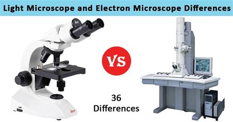
The core difference between light and electron microscopes lies in their method of illumination. Light microscopes, as their name suggests, use visible light to illuminate the specimen. This light passes through a series of lenses to magnify the image, allowing us to observe the specimen's structure. Electron microscopes, on the other hand, employ a beam of electrons instead of light. The electrons, having a much shorter wavelength than visible light, enable far greater resolution and magnification.
Light Microscopy: A Closer Look
Light microscopy, a staple in biology and other scientific disciplines, relies on the interaction of light with the specimen. Various techniques exist, each with its unique approach:
- Bright-field microscopy: This is the most basic type, where light passes directly through the specimen. Staining is often required to enhance contrast and visibility.
- Dark-field microscopy: Here, light is directed at an angle, illuminating only the edges of the specimen, creating a bright object against a dark background. This is beneficial for observing unstained, transparent specimens.
- Phase-contrast microscopy: This technique enhances contrast by exploiting differences in the refractive index of various parts of the specimen. It is ideal for observing living cells without staining.
- Fluorescence microscopy: This advanced technique employs fluorescent dyes or proteins that emit light at a specific wavelength when excited by light of a different wavelength. This enables highly specific labeling and visualization of cellular structures.
- Confocal microscopy: A sophisticated variant of fluorescence microscopy that uses lasers and pinholes to eliminate out-of-focus light, resulting in sharp, three-dimensional images.
Electron Microscopy: Delving into the Ultrastructure
Electron microscopy leverages the properties of electrons to achieve significantly higher resolution. The process involves:
- Transmission Electron Microscopy (TEM): In TEM, a beam of electrons is transmitted through an ultra-thin specimen. The electrons interact with the specimen, and the resulting pattern is projected onto a screen or detector, producing a highly magnified image. This technique reveals intricate internal details of cells and other structures.
- Scanning Electron Microscopy (SEM): Unlike TEM, SEM scans the surface of a specimen with a focused beam of electrons. The electrons interact with the surface atoms, generating signals that are used to create a three-dimensional image. SEM is particularly useful for visualizing surface topography and texture.
- Scanning Transmission Electron Microscopy (STEM): This technique combines aspects of both TEM and SEM, providing high-resolution images of both internal structure and surface features.
A Head-to-Head Comparison: Key Differences and Similarities
The following table summarizes the key differences and similarities between light and electron microscopes:
| Feature | Light Microscope | Electron Microscope |
|---|---|---|
| Illumination | Visible light | Beam of electrons |
| Wavelength | Relatively long (400-700 nm) | Extremely short (0.004 nm for electrons) |
| Magnification | Up to 1500x | Up to 1,000,000x |
| Resolution | Limited by wavelength (around 200 nm) | Much higher (0.1 nm or better) |
| Sample Prep | Relatively simple (staining, mounting) | Complex (thin sectioning, fixation, coating) |
| Cost | Relatively inexpensive | Very expensive |
| Specimen type | Living or fixed specimens | Usually fixed, dehydrated specimens |
| Vacuum Required | No | Yes (for electron microscopes) |
| Image type | 2D or (with advanced techniques) 3D | Primarily 2D (SEM offers 3D surface view) |
Advantages and Disadvantages: Choosing the Right Tool
The choice between a light microscope and an electron microscope depends heavily on the specific application and the level of detail required.
Light Microscopy: Advantages & Disadvantages
Advantages:
- Relatively inexpensive and easy to use: Light microscopes are accessible and require less specialized training.
- Can observe living specimens: Many light microscopy techniques allow for the observation of living cells and their dynamic processes.
- Sample preparation is simpler: The preparation methods are less time-consuming and less complex.
- Versatile techniques: Offers a range of techniques for different applications (bright-field, dark-field, phase-contrast, fluorescence, confocal).
Disadvantages:
- Lower resolution: Limited by the wavelength of light, resulting in lower resolution compared to electron microscopes.
- Limited magnification: Cannot achieve the high magnifications possible with electron microscopes.
- Staining can affect specimens: Staining techniques can sometimes alter or damage the specimen.
Electron Microscopy: Advantages & Disadvantages
Advantages:
- High resolution and magnification: Enables visualization of extremely fine details, revealing structures invisible to light microscopes.
- Detailed structural information: Provides detailed insights into the ultrastructure of cells, tissues, and materials.
- Versatile techniques: Different types of electron microscopy (TEM, SEM, STEM) cater to various needs.
Disadvantages:
- High cost and complexity: Electron microscopes are significantly more expensive and require specialized training to operate.
- Specimen preparation is complex and time-consuming: Preparing samples for electron microscopy requires meticulous techniques that can be difficult to master.
- Vacuum environment: The need for a vacuum environment means that live specimens cannot be observed.
- Can induce artifacts: The preparation process may introduce artifacts that can distort the image.
- Limited penetration depth in SEM: SEM primarily shows surface features, with limited information about deeper structures.
Applications of Light and Electron Microscopes
Both light and electron microscopes are indispensable tools across various scientific disciplines.
Applications of Light Microscopy
- Biology: Observing living cells, cell division, microorganisms, and tissue samples.
- Medicine: Diagnosing diseases, analyzing blood samples, examining tissue biopsies.
- Material science: Analyzing the microstructure of materials.
- Forensic science: Analyzing evidence such as fibers and hairs.
- Education: Teaching basic biological principles and microscopy techniques.
Applications of Electron Microscopy
- Materials science: Characterizing the microstructure of materials, studying defects and fractures.
- Nanotechnology: Visualizing and analyzing nanostructures and devices.
- Biology: Studying the ultrastructure of cells, organelles, and viruses.
- Medicine: Diagnosing diseases, analyzing tissues at a high resolution.
- Environmental science: Analyzing pollutants and microorganisms in the environment.
The Future of Microscopy: Beyond Light and Electrons
Microscopy continues to evolve, with new techniques constantly emerging. Super-resolution microscopy, for instance, pushes the boundaries of light microscopy, achieving resolutions far beyond the diffraction limit of light. Cryo-electron microscopy (cryo-EM) has revolutionized structural biology, enabling high-resolution imaging of biomolecules in their near-native state. These advanced techniques are expanding our ability to visualize the incredibly small, promising even more groundbreaking discoveries in the future.
Conclusion: Synergy and Specialization
While light and electron microscopes offer distinct advantages and disadvantages, they are not mutually exclusive. Often, researchers utilize both types of microscopy in a complementary manner. Light microscopy provides a broader overview, while electron microscopy reveals the intricate details. This synergistic approach unlocks a more comprehensive understanding of the microscopic world, driving advancements in various fields of science and technology. The choice ultimately depends on the specific research question, desired level of detail, and available resources. Understanding the strengths and limitations of both techniques is crucial for any researcher working in microscopy-based fields.
Latest Posts
Latest Posts
-
Difference Between Spongy And Compact Bone
Apr 04, 2025
-
D How Is The Energy Produced By Respiration Stored
Apr 04, 2025
-
The Functional And Structural Unit Of The Kidneys Is The
Apr 04, 2025
-
To Pour Water On Calcium Oxide
Apr 04, 2025
-
Evaluate The Trigonometric Function At The Quadrantal Angle
Apr 04, 2025
Related Post
Thank you for visiting our website which covers about Comparison Between Light Microscope And Electron Microscope . We hope the information provided has been useful to you. Feel free to contact us if you have any questions or need further assistance. See you next time and don't miss to bookmark.
