Which Phase Of Mitosis Is Shown In The Image
Juapaving
Mar 12, 2025 · 6 min read
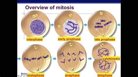
Table of Contents
Which Phase of Mitosis is Shown in the Image? A Comprehensive Guide
Determining the specific phase of mitosis depicted in a microscopic image requires careful observation of several key features. Mitosis, the process of cell division resulting in two identical daughter cells, is a complex sequence of events. Understanding these phases – prophase, prometaphase, metaphase, anaphase, and telophase – is crucial for accurate identification. This guide will provide a detailed explanation of each phase, helping you confidently identify the mitotic phase shown in any given image.
Understanding the Stages of Mitosis
Mitosis is a continuous process, but for ease of understanding, it's divided into distinct phases based on observable changes within the cell. Let's examine each one:
1. Prophase: The Initial Stage of Chromosomal Condensation
Prophase marks the beginning of mitosis. Several key characteristics distinguish this phase:
- Chromatin Condensation: The chromatin, the loosely organized DNA and protein complex within the nucleus, begins to condense into visible, distinct chromosomes. Each chromosome consists of two identical sister chromatids joined at the centromere. This condensation is crucial for efficient segregation during later stages.
- Nuclear Envelope Breakdown: The nuclear envelope, the membrane surrounding the nucleus, begins to fragment and disintegrate. This allows the chromosomes to access the cytoplasm and interact with the mitotic spindle.
- Spindle Formation: The mitotic spindle, a complex structure composed of microtubules, begins to form. These microtubules originate from the centrosomes, which have duplicated earlier in the cell cycle and are migrating to opposite poles of the cell. The spindle fibers will play a crucial role in chromosome movement.
- Nucleolus Disappearance: The nucleolus, a dense region within the nucleus responsible for ribosome synthesis, disappears. This is a consequence of the overall disruption of the nuclear structure.
Identifying Prophase in an Image: Look for condensed chromosomes within a still-intact or partially disintegrating nuclear envelope. The chromosomes will appear as thick, rod-like structures. The mitotic spindle might be partially formed, but not yet fully organized.
2. Prometaphase: Chromosomes Attach to the Spindle Apparatus
Prometaphase is a transitional phase between prophase and metaphase. The defining feature is the attachment of chromosomes to the mitotic spindle:
- Microtubule Attachment: Spindle microtubules extend from the centrosomes and attach to the kinetochores, specialized protein structures located at the centromeres of each chromosome. This attachment is crucial for accurate chromosome segregation. Each sister chromatid has its own kinetochore.
- Chromosome Movement: The chromosomes begin to move towards the metaphase plate, an imaginary plane equidistant from the two spindle poles. This movement is a result of the dynamic interactions between the kinetochore microtubules and the motor proteins associated with the kinetochores.
- Nuclear Envelope Complete Disintegration: Any remaining fragments of the nuclear envelope completely disappear during prometaphase.
Identifying Prometaphase in an Image: Look for chromosomes with attached spindle fibers, actively moving towards the cell's center. The chromosomes are still fairly condensed, but might show some alignment towards a central plane. The absence of a nuclear envelope is key here.
3. Metaphase: Chromosomes Align at the Metaphase Plate
Metaphase is characterized by the precise alignment of chromosomes at the metaphase plate:
- Chromosome Alignment: All chromosomes are aligned at the metaphase plate, with their centromeres positioned exactly in the middle. This alignment is crucial for ensuring that each daughter cell receives one copy of each chromosome.
- Sister Chromatid Connection: Sister chromatids remain connected at the centromere. The tension generated by the opposing forces of the kinetochore microtubules ensures the proper alignment.
- Spindle Checkpoint: A critical checkpoint in the cell cycle occurs during metaphase. The cell ensures that all chromosomes are correctly attached to the spindle before proceeding to anaphase. This checkpoint prevents aneuploidy (abnormal chromosome numbers) in daughter cells.
Identifying Metaphase in an Image: Observe the chromosomes precisely aligned along the metaphase plate. Sister chromatids are clearly visible and attached at the centromere. The spindle fibers appear clearly attached to the kinetochores. This is a very organized and symmetrical arrangement.
4. Anaphase: Sister Chromatids Separate and Migrate
Anaphase is characterized by the separation of sister chromatids and their movement to opposite poles of the cell:
- Sister Chromatid Separation: The centromeres divide, and the sister chromatids separate, becoming individual chromosomes. This separation is triggered by the activation of separase, an enzyme that cleaves the protein complex holding sister chromatids together.
- Chromosome Movement: The individual chromosomes are pulled towards the opposite poles of the cell by the shortening of the kinetochore microtubules. This movement is powered by motor proteins and the depolymerization of microtubules.
- Spindle Elongation: The spindle itself elongates, further separating the chromosomes.
Identifying Anaphase in an Image: Look for individual chromosomes moving towards opposite poles of the cell. The chromosomes appear "V"-shaped as they are pulled by the kinetochore microtubules. The distance between the chromosomes and the poles will progressively decrease as the phase progresses.
5. Telophase: Chromosomes Decondense and Nuclear Envelope Reforms
Telophase marks the final stage of mitosis:
- Chromosome Decondensation: The chromosomes begin to decondense, reverting to their less-condensed chromatin form.
- Nuclear Envelope Reformation: A new nuclear envelope forms around each set of chromosomes at the poles of the cell.
- Nucleolus Reappearance: The nucleolus reappears within each new nucleus.
- Spindle Disassembly: The mitotic spindle disassembles.
Identifying Telophase in an Image: Look for decondensed chromosomes within newly formed nuclear envelopes. The spindle will be disappearing or gone. The cell is preparing for cytokinesis, the division of the cytoplasm.
Cytokinesis: Completing Cell Division
Cytokinesis is not technically part of mitosis but is the final step in the cell cycle, following telophase. It involves the division of the cytoplasm, resulting in two separate daughter cells, each with a complete set of chromosomes. In animal cells, cytokinesis is achieved by a contractile ring of actin filaments that pinches the cell in two. In plant cells, a cell plate forms between the two daughter nuclei, eventually developing into a new cell wall.
Analyzing Microscopic Images: Tips for Accurate Identification
To accurately determine the phase of mitosis shown in an image, consider the following:
- Chromosome Condensation: The degree of condensation increases from prophase to metaphase and then decreases in telophase.
- Nuclear Envelope Integrity: The nuclear envelope is intact in prophase, fragments during prometaphase, and is completely absent in metaphase and anaphase. It reforms in telophase.
- Spindle Apparatus: The spindle forms and becomes more organized throughout mitosis, reaching its peak complexity in metaphase and anaphase, and then disassembles in telophase.
- Chromosome Alignment: Chromosomes align at the metaphase plate in metaphase. In anaphase, they are separated and moving towards the poles.
- Sister Chromatid Separation: Sister chromatids separate in anaphase.
By carefully observing these features in a microscopic image, you can accurately determine which phase of mitosis is depicted. Remember that the transition between phases is gradual, and images might show features intermediate between two consecutive phases. Consider the predominant characteristics to make the most accurate determination. Practice with various images to sharpen your skills in identifying the different stages of mitosis. The more images you analyze, the better your ability will become to differentiate between the subtle differences in each phase. Careful observation and a methodical approach are key to mastering this essential skill in cell biology.
Latest Posts
Latest Posts
-
Sample Letter Of Refund Payment To Customer
May 09, 2025
-
What Is The Source Of Oxygen Released During Photosynthesis
May 09, 2025
-
How Many Centimeters Is 13 Inches
May 09, 2025
-
Find The Number Of Edges On This Solid
May 09, 2025
-
Are Hydrogen Bonds Weaker Than Covalent Bonds
May 09, 2025
Related Post
Thank you for visiting our website which covers about Which Phase Of Mitosis Is Shown In The Image . We hope the information provided has been useful to you. Feel free to contact us if you have any questions or need further assistance. See you next time and don't miss to bookmark.