Where Are The Cardiac Vasomotor And Respiratory Centers Found
Juapaving
Mar 12, 2025 · 6 min read
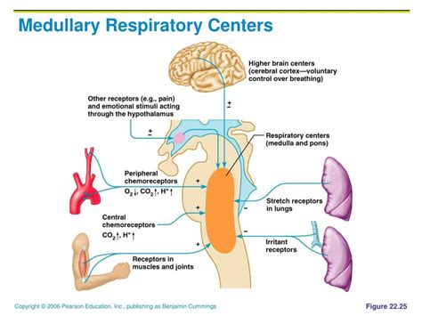
Table of Contents
Where Are the Cardiac, Vasomotor, and Respiratory Centers Found?
The human body is a marvel of intricate systems working in perfect harmony. At the heart of this orchestration lies the brainstem, a crucial structure responsible for many essential autonomic functions. Among these, the regulation of cardiovascular function (cardiac and vasomotor centers) and breathing (respiratory center) are paramount for survival. Understanding the precise location and interplay of these centers is fundamental to comprehending physiological processes and diagnosing neurological disorders. This article delves deep into the neuroanatomy of these vital centers, exploring their location, function, and interactions.
Locating the Vital Control Centers in the Brainstem
The brainstem, the stalk-like structure connecting the cerebrum to the spinal cord, houses several crucial nuclei that coordinate vital functions. The cardiac, vasomotor, and respiratory centers are not discrete, anatomically separate entities but rather complex networks of interconnected neurons residing within specific brainstem regions. Their precise boundaries are somewhat fuzzy, with significant overlap and interaction.
The Medulla Oblongata: The Heart of Autonomic Control
The medulla oblongata, the most caudal part of the brainstem, plays the central role in housing these vital centers. Its strategic position ensures direct communication with the spinal cord and other brainstem regions. Within the medulla, we find the key components:
1. The Cardiac Center: Regulating Heart Rate
The cardiac center is not a single, well-defined nucleus but a collection of neurons scattered within the medulla. It's primarily located in the dorsal medulla, in close proximity to the solitary nucleus (NTS), a crucial integration center for visceral sensory input. The cardiac center is further divided into two functionally distinct components:
-
Cardioinhibitory Center: This center primarily influences parasympathetic activity, slowing down the heart rate. It achieves this by sending signals via the vagus nerve (CN X) to the sinoatrial (SA) node, the heart's natural pacemaker. Increased activity in this center leads to bradycardia (slow heart rate).
-
Cardioacceleratory Center: This center modulates sympathetic activity, increasing the heart rate and contractility. Signals are transmitted via the sympathetic nervous system, specifically the cervical and upper thoracic spinal cord, to the SA node and myocardium. Increased activity here leads to tachycardia (rapid heart rate).
The balance between the cardioinhibitory and cardioacceleratory centers determines the overall heart rate and contractility.
2. The Vasomotor Center: Controlling Blood Vessel Tone
Adjacent to the cardiac center, also within the dorsal medulla, lies the vasomotor center. This is similarly a collection of neurons, not a single, sharply defined structure. Its primary function is to regulate blood vessel tone, impacting blood pressure. Like the cardiac center, it also operates through sympathetic and parasympathetic pathways:
-
Sympathetic Pathways: The vasomotor center primarily utilizes sympathetic pathways to control blood vessel constriction (vasoconstriction). Signals travel down the spinal cord and out to the peripheral blood vessels, causing them to narrow, increasing peripheral resistance and blood pressure. This is mediated primarily through the release of norepinephrine.
-
Parasympathetic Pathways: The influence of the parasympathetic system on blood vessel tone is less direct and primarily affects specific vascular beds, such as those in the salivary glands and digestive tract. Parasympathetic stimulation generally causes vasodilation.
The vasomotor center constantly monitors blood pressure through baroreceptors and chemoreceptors located in the circulatory system. These receptors relay information back to the center, allowing for continuous feedback and fine-tuning of blood vessel tone to maintain blood pressure within a narrow range.
3. The Respiratory Center: Orchestrating Breathing
The respiratory center, arguably the most crucial of the three, is also located in the medulla, but spans a larger area than the cardiac and vasomotor centers. It comprises several interconnected groups of neurons:
-
Dorsal Respiratory Group (DRG): Situated in the dorsal medulla, the DRG is primarily involved in initiating inspiration (breathing in). It receives sensory input from peripheral chemoreceptors and sends signals to the inspiratory muscles (diaphragm and external intercostals).
-
Ventral Respiratory Group (VRG): Located in the ventral medulla, the VRG is involved in both inspiration and expiration. During quiet breathing, its activity is minimal. However, during forceful breathing (e.g., exercise), it becomes crucial in coordinating both inspiratory and expiratory muscles.
-
Pneumotaxic Center (Pontine Respiratory Group): While not directly in the medulla, the pneumotaxic center, located in the pons (the brain region superior to the medulla), plays a critical role in regulating the respiratory rhythm. It primarily limits the duration of inspiration, contributing to the smooth transition between inspiration and expiration.
The interplay between these medullary and pontine groups finely tunes the respiratory rhythm, ensuring efficient gas exchange. They respond to changes in blood gas levels (oxygen, carbon dioxide) and pH, adjusting breathing rate and depth accordingly.
Interaction and Integration of the Centers
It's crucial to understand that these three centers—cardiac, vasomotor, and respiratory—do not operate in isolation. They constantly interact and influence one another, maintaining a finely tuned homeostatic balance. For example:
-
Cardiovascular and Respiratory Interactions: Changes in blood gas levels (e.g., increased carbon dioxide) stimulate the respiratory center, leading to increased breathing rate. This, in turn, can influence blood pressure by altering venous return to the heart.
-
Vasomotor and Cardiac Interactions: The vasomotor center's regulation of blood pressure directly impacts the workload of the heart. Increased blood pressure increases cardiac work, potentially triggering adjustments in heart rate and contractility mediated by the cardiac center.
-
Integrated Response to Stress: During stress, the sympathetic nervous system is activated, leading to simultaneous increases in heart rate (via the cardiac center), blood pressure (via the vasomotor center), and respiratory rate (via the respiratory center). This integrated response prepares the body for "fight or flight."
Clinical Significance
Understanding the location and function of these vital centers is critical in clinical practice. Damage to the medulla, whether from trauma, stroke, or other neurological conditions, can severely disrupt these autonomic functions, leading to potentially fatal consequences. Conditions affecting these centers can manifest as:
-
Bradycardia or Tachycardia: Imbalances in the cardiac center can result in abnormally slow or fast heart rates.
-
Hypertension or Hypotension: Dysregulation of the vasomotor center can lead to dangerously high or low blood pressure.
-
Respiratory Distress or Failure: Damage to the respiratory center can severely impair breathing, requiring respiratory support.
Conclusion
The cardiac, vasomotor, and respiratory centers are complex networks of neurons residing primarily within the medulla oblongata, forming the foundation of autonomic control. Their precise location, intricate interplay, and integrated responses to various stimuli are crucial for maintaining homeostasis and survival. Understanding the neuroanatomy and functional interactions of these centers provides invaluable insight into physiological processes and is essential for diagnosing and managing neurological conditions that affect vital autonomic functions. Further research continues to unravel the intricacies of these vital brainstem nuclei, continually refining our understanding of their crucial role in maintaining life.
Latest Posts
Latest Posts
-
How Many Degrees Are In A Half Circle
May 09, 2025
-
What Force Keeps The Planets In Orbit Around The Sun
May 09, 2025
-
What Are The Products Of This Chemical Reaction
May 09, 2025
-
What Are Some Examples Of A Screw
May 09, 2025
-
What Planet Is Known As The Morning Star
May 09, 2025
Related Post
Thank you for visiting our website which covers about Where Are The Cardiac Vasomotor And Respiratory Centers Found . We hope the information provided has been useful to you. Feel free to contact us if you have any questions or need further assistance. See you next time and don't miss to bookmark.