This Image Shows The Tertiary Structure Of A Protein Segment
Juapaving
Jun 01, 2025 · 6 min read
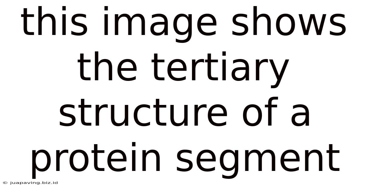
Table of Contents
Decoding the Tertiary Structure: A Deep Dive into Protein Folding
This image showcases the tertiary structure of a protein segment, a fascinating and crucial aspect of biochemistry. Understanding this complex three-dimensional arrangement is essential for comprehending protein function and the implications of structural misfolding in various diseases. This article will delve into the intricacies of tertiary protein structure, exploring the forces driving its formation, the diverse structural motifs involved, and the significant consequences of structural alterations.
Understanding Protein Structure: A Hierarchical Approach
Proteins are the workhorses of the cell, performing a vast array of functions, from catalyzing biochemical reactions to providing structural support. Their ability to perform these diverse tasks is intimately linked to their complex three-dimensional structures, which are hierarchically organized into four levels:
1. Primary Structure: The Linear Sequence
The primary structure is the fundamental building block, representing the linear sequence of amino acids linked together by peptide bonds. This sequence is dictated by the genetic code encoded in DNA. The specific order of amino acids is critical, as it determines all subsequent levels of protein structure. A single amino acid substitution can drastically alter the protein's function.
2. Secondary Structure: Local Folding Patterns
Once synthesized, the polypeptide chain begins to fold into local secondary structures. These are stabilized by hydrogen bonds between the backbone atoms of the amino acids. Common secondary structures include:
- α-helices: Right-handed coiled structures stabilized by hydrogen bonds between the carbonyl oxygen of one amino acid and the amide hydrogen of an amino acid four residues down the chain.
- β-sheets: Extended structures formed by hydrogen bonds between adjacent polypeptide strands. These strands can be parallel or anti-parallel, depending on the orientation of the amino acid chains.
- Loops and turns: Irregular regions connecting α-helices and β-sheets, often crucial for protein function and interaction with other molecules.
3. Tertiary Structure: The 3D Arrangement
The tertiary structure refers to the overall three-dimensional arrangement of a polypeptide chain, including all its secondary structure elements. This is a crucial level of organization as it dictates the protein's function. The tertiary structure is stabilized by a variety of weak and strong interactions, including:
- Disulfide bonds: Covalent bonds formed between cysteine residues, creating strong cross-links within the protein.
- Hydrogen bonds: These form between various polar groups within the protein, contributing to the overall stability.
- Ionic interactions (salt bridges): Electrostatic interactions between charged amino acid side chains.
- Hydrophobic interactions: Nonpolar amino acid side chains cluster together in the protein's core, minimizing their contact with water.
- Van der Waals forces: Weak attractive forces between atoms in close proximity.
The interplay of these forces determines the precise folding pathway and the final three-dimensional structure of the protein. This process is often complex and can involve chaperone proteins that assist in proper folding.
4. Quaternary Structure: Multiple Polypeptide Chains
Some proteins consist of multiple polypeptide chains, each with its own tertiary structure. The arrangement of these individual subunits forms the quaternary structure. The interactions between subunits are similar to those stabilizing the tertiary structure, including hydrogen bonds, ionic interactions, and hydrophobic interactions. Examples of proteins with quaternary structure include hemoglobin and many enzymes.
Factors Influencing Tertiary Structure
The tertiary structure of a protein is not arbitrary; it's a consequence of a delicate balance between various factors. Understanding these factors is crucial to understanding the protein's function and potential vulnerabilities.
Amino Acid Sequence: The Blueprint
The primary amino acid sequence dictates the tertiary structure. The unique sequence determines the location of hydrophobic and hydrophilic residues, influencing how the protein folds to minimize unfavorable interactions with water. The presence of cysteine residues, capable of forming disulfide bonds, also significantly affects the final structure.
Environmental Conditions: The Shaping Hand
Environmental factors such as temperature, pH, and the presence of ions can drastically affect protein folding and stability. Changes in these conditions can disrupt weak interactions, leading to denaturation – the unfolding of the protein. This can lead to loss of function or even aggregation of misfolded proteins, which is implicated in several diseases.
Chaperone Proteins: The Guiding Hand
Chaperone proteins play a critical role in guiding the proper folding of nascent polypeptides. They prevent aggregation and ensure that proteins fold into their functional conformation. They achieve this by providing a protected environment where folding can proceed without interference from other molecules.
Common Tertiary Structure Motifs
While the tertiary structure of each protein is unique, some recurring structural motifs are frequently observed. These motifs represent stable folding patterns that are favored due to their thermodynamic stability.
α/β barrels: Combining Helices and Sheets
This motif combines α-helices and β-sheets in a barrel-like structure. The β-sheets form the barrel's walls, and the α-helices connect the strands, stabilizing the structure. This motif is prevalent in enzymes involved in various metabolic processes.
Zinc finger motifs: Zinc Coordination
Zinc finger motifs are characterized by a zinc ion coordinated by cysteine and histidine residues. These motifs often bind DNA, playing a crucial role in gene regulation.
Rossmann folds: β-α-β motifs
This motif consists of alternating β-strands and α-helices, often involved in binding nucleotide cofactors such as NAD+ and FAD. It's common in enzymes involved in redox reactions.
SH2 domains: Phosphotyrosine recognition
These domains specifically recognize phosphotyrosine residues, playing a crucial role in signaling pathways. They contribute to protein-protein interactions and are vital in cell regulation.
Consequences of Tertiary Structure Misfolding
The proper folding of proteins is essential for their function. Misfolding can lead to a range of consequences, often resulting in disease. This can stem from genetic mutations altering the amino acid sequence, environmental stresses disrupting weak interactions, or deficiencies in chaperone proteins.
Amyloid diseases: Protein Aggregation
Misfolded proteins can aggregate, forming amyloid fibrils – insoluble protein deposits that accumulate in tissues. These aggregates are associated with various neurodegenerative diseases, including Alzheimer's disease, Parkinson's disease, and Huntington's disease.
Cancer: Dysfunctional Proteins
Misfolded proteins can disrupt cellular processes, leading to uncontrolled cell growth and cancer. Mutations in tumor suppressor genes or oncogenes can lead to the production of misfolded proteins that contribute to cancer development.
Prion diseases: Infectious Misfolding
Prion diseases, such as Creutzfeldt-Jakob disease, are caused by the misfolding of prion proteins. The misfolded form can then induce the misfolding of other prion proteins, leading to a chain reaction of misfolding and aggregation.
Conclusion: The Intricate World of Protein Structure
The tertiary structure of a protein is a testament to the exquisite complexity of biological systems. Its intricate three-dimensional arrangement, shaped by the interplay of various forces and environmental factors, is essential for protein function. Understanding the forces that drive protein folding and the consequences of misfolding is critical for advancing our understanding of health and disease, potentially leading to novel therapeutic interventions for protein misfolding-related diseases. Further research into the intricacies of protein structure continues to unlock the secrets of life itself. This exploration into the tertiary structure is far from complete, and future advancements in imaging techniques, computational modeling, and biochemical assays are poised to reveal even more about this fascinating aspect of biological systems. The journey to fully understanding the intricacies of protein folding and its impact remains a challenging yet incredibly rewarding pursuit.
Latest Posts
Related Post
Thank you for visiting our website which covers about This Image Shows The Tertiary Structure Of A Protein Segment . We hope the information provided has been useful to you. Feel free to contact us if you have any questions or need further assistance. See you next time and don't miss to bookmark.