The Study Of Tissue Is Known As
Juapaving
Apr 05, 2025 · 7 min read

Table of Contents
The Study of Tissue is Known As: A Deep Dive into Histology
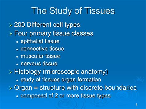
The study of tissue is known as histology. Histology is a crucial branch of biology that bridges the gap between the macroscopic world we see and the microscopic world that governs the function of living organisms. It involves the detailed examination of tissues, their structure, composition, and function, providing invaluable insights into health, disease, and the intricate workings of the human body and beyond. This comprehensive exploration delves into the fascinating field of histology, exploring its techniques, applications, and importance in various scientific disciplines.
What is Histology?
Histology, derived from the Greek words "histos" (tissue) and "logos" (study), is the microscopic study of the structure and function of tissues. Tissues are groups of cells that have similar structures and work together to perform specific functions. Understanding the organization and arrangement of these cells within tissues is fundamental to comprehending the overall structure and function of organs and organ systems. Histology isn't simply about identifying cell types; it's about deciphering the complex interplay between cells, the extracellular matrix (ECM), and the overall tissue architecture. This intricate relationship dictates the tissue's mechanical properties, its responsiveness to stimuli, and its susceptibility to disease.
The Importance of Histology
The importance of histology extends far beyond academic curiosity. It plays a critical role in:
1. Disease Diagnosis:
Pathology, a cornerstone of modern medicine, relies heavily on histological analysis. By examining tissue samples (biopsies), pathologists can identify cancerous cells, inflammatory processes, infectious agents, and other abnormalities. The accurate diagnosis of diseases like cancer, autoimmune disorders, and infectious diseases depends crucially on the precise identification of tissue changes at the microscopic level. The ability to distinguish between normal and abnormal tissue morphology is paramount in guiding treatment decisions and predicting prognosis.
2. Research and Development:
Histology is indispensable in scientific research across various fields. Researchers utilize histological techniques to investigate:
- Developmental biology: Studying the formation and differentiation of tissues during embryonic development.
- Pharmacology: Evaluating the effects of drugs and other therapeutic agents on tissues.
- Toxicology: Assessing the toxicity of various substances on cellular and tissue levels.
- Regenerative medicine: Investigating tissue regeneration and repair mechanisms.
- Forensic science: Identifying and analyzing tissue samples in forensic investigations.
3. Understanding Physiological Processes:
Histology provides critical insights into the physiological processes occurring within the body. By examining the structure and organization of tissues, researchers can better understand how these processes work at a cellular and tissue level. For example, histological analysis can reveal the intricate mechanisms of nutrient absorption in the intestines, the precise arrangement of cells in the nervous system allowing for signal transmission, or the complex processes involved in muscle contraction.
Histological Techniques: Preparing Tissues for Examination
Examining tissues at a microscopic level requires careful preparation. The process typically involves several crucial steps:
1. Tissue Fixation:
The first step involves tissue fixation, which preserves the tissue's structure and prevents degradation. Common fixatives include formalin, which cross-links proteins and stabilizes the tissue. The choice of fixative depends on the specific tissue and the type of analysis planned.
2. Tissue Processing:
After fixation, the tissue undergoes processing, a series of steps designed to prepare it for embedding. This often involves dehydration (removing water) using increasing concentrations of alcohol, followed by clearing with solvents like xylene to remove the alcohol.
3. Embedding:
The processed tissue is then embedded in a medium, usually paraffin wax, which provides support during sectioning. The wax-embedded tissue block is allowed to solidify.
4. Sectioning:
A microtome is used to create very thin sections (typically 3-5 micrometers thick) of the embedded tissue. These thin sections are essential for allowing light to pass through the tissue during microscopic examination.
5. Staining:
Tissue sections are stained to enhance contrast and highlight specific cellular components. Hematoxylin and eosin (H&E) staining is a widely used routine stain. Hematoxylin stains nuclei blue/purple, while eosin stains the cytoplasm and extracellular matrix pink/red. Other specialized stains can highlight specific structures like collagen fibers, elastic fibers, or microorganisms. Immunohistochemistry (IHC) and in situ hybridization (ISH) techniques use antibodies or labeled probes to identify specific molecules within tissues, providing more detailed information about cellular composition and function.
6. Microscopy:
Finally, the stained tissue sections are examined using a light microscope. The resolution of a light microscope allows visualization of cellular structures down to approximately 0.2 micrometers. For higher resolution, electron microscopy can be employed. Electron microscopy uses beams of electrons instead of light and offers significantly higher magnification and resolution, allowing the visualization of subcellular structures like organelles and macromolecules.
Types of Tissues: A Histological Overview
The human body is composed of four primary tissue types:
1. Epithelial Tissue:
Epithelial tissues cover body surfaces, line cavities and organs, and form glands. They are characterized by closely packed cells with minimal extracellular matrix. Epithelial tissues are classified based on cell shape (squamous, cuboidal, columnar) and the number of layers (simple, stratified, pseudostratified). Examples include the epidermis of the skin, the lining of the digestive tract, and the glandular tissue of the endocrine system. Their functions include protection, secretion, absorption, and excretion.
2. Connective Tissue:
Connective tissues support, connect, and separate different tissues and organs. They are characterized by abundant extracellular matrix, which is composed of fibers (collagen, elastic, reticular) and ground substance. Types of connective tissues include loose connective tissue, dense connective tissue, adipose tissue, cartilage, bone, and blood. Their functions vary widely, including structural support, nutrient storage, and immune defense.
3. Muscle Tissue:
Muscle tissues are responsible for movement. There are three types of muscle tissue: skeletal muscle, smooth muscle, and cardiac muscle. Skeletal muscle is responsible for voluntary movement, smooth muscle for involuntary movements in internal organs, and cardiac muscle for the contraction of the heart. Their distinct microscopic structures reflect their different functional roles.
4. Nervous Tissue:
Nervous tissue is specialized for the rapid transmission of electrical signals. It is composed of neurons, which are responsible for generating and transmitting nerve impulses, and glial cells, which support and protect neurons. The arrangement of neurons and glial cells within the nervous system is crucial for information processing and communication throughout the body.
Histology's Expanding Horizons: Advanced Techniques and Applications
The field of histology is constantly evolving, with new techniques and applications emerging regularly.
1. Immunohistochemistry (IHC):
IHC utilizes antibodies to detect specific proteins within tissues. This technique is invaluable in diagnosing cancers, identifying infectious agents, and studying the expression of specific genes and proteins.
2. In Situ Hybridization (ISH):
ISH uses labeled probes to detect specific DNA or RNA sequences within tissues. This allows for the identification of specific genes and their expression patterns within cells and tissues.
3. Confocal Microscopy:
Confocal microscopy is a powerful technique that allows for the creation of high-resolution images of three-dimensional structures. This technique is particularly useful for studying the complex architecture of tissues and the interactions between different cell types.
4. Digital Histopathology:
Digital histopathology involves the digitization of microscopic images, allowing for remote viewing, analysis, and sharing of histological data. This fosters collaboration, improves diagnostic efficiency, and allows for the development of artificial intelligence-based diagnostic tools.
5. 3D Histology and Tissue Engineering:
Advances in imaging and computational modeling are enabling the creation of three-dimensional models of tissues, furthering our understanding of tissue architecture and paving the way for advances in tissue engineering and regenerative medicine. The ability to reconstruct tissue structures in 3D provides an unprecedented level of detail for analyzing tissue organization and function.
Conclusion: The Enduring Significance of Histology
Histology remains a cornerstone of biological and medical research, providing invaluable insights into the structure, function, and pathology of tissues. From diagnosing diseases to advancing our understanding of fundamental biological processes, histology's impact is far-reaching. As technology continues to advance, so too will the capabilities of histology, opening new avenues for research and offering new possibilities for improving human health and well-being. The intricate world revealed by histological techniques continues to captivate researchers and healthcare professionals alike, underscoring its enduring significance in the scientific landscape. The detailed study of tissues continues to unravel the mysteries of life itself and to push the boundaries of medical innovation.
Latest Posts
Latest Posts
-
Examples Of Abiotic Factors And Biotic Factors
Apr 06, 2025
-
For Every Action There Is An Equal Opposite Reaction
Apr 06, 2025
-
A Comparison Of Two Similar Quantities Using Division
Apr 06, 2025
-
Round 0 234 To The Nearest Hundredth
Apr 06, 2025
-
An Inch Is What Fraction Of A Yard
Apr 06, 2025
Related Post
Thank you for visiting our website which covers about The Study Of Tissue Is Known As . We hope the information provided has been useful to you. Feel free to contact us if you have any questions or need further assistance. See you next time and don't miss to bookmark.
