The Inspiratory And Expiratory Centers Are Located In The
Juapaving
Mar 13, 2025 · 6 min read
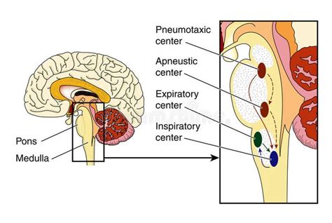
Table of Contents
The Inspiratory and Expiratory Centers: Location, Function, and Clinical Significance
The rhythmic process of breathing, essential for life, is orchestrated by a complex interplay of neural circuits located primarily within the brainstem. Understanding the location and function of these centers, specifically the inspiratory and expiratory centers, is crucial for comprehending respiratory physiology and various respiratory disorders. This article will delve deep into the precise location, the intricate mechanisms of these centers, and their clinical significance.
Location of the Respiratory Centers
The respiratory centers aren't confined to a single, easily definable area. Instead, they're a network of interconnected neurons distributed throughout the brainstem, primarily within the medulla oblongata and pons. This network involves several key areas:
Medulla Oblongata: The Primary Control Center
The medulla oblongata houses the crucial dorsal respiratory group (DRG) and the ventral respiratory group (VRG). These groups are not discrete, sharply defined structures, but rather collections of neurons with overlapping functions.
-
Dorsal Respiratory Group (DRG): Located in the nucleus tractus solitarius (NTS), the DRG is considered the primary rhythm generator for inspiration. Its neurons fire rhythmically, initiating inspiration by sending signals to the inspiratory muscles, primarily the diaphragm. The DRG receives sensory input from various peripheral chemoreceptors and mechanoreceptors, helping regulate breathing based on the body's needs. Think of the DRG as the pacemaker setting the basic rhythm of breathing.
-
Ventral Respiratory Group (VRG): Situated more ventrally in the medulla, the VRG is active during both inspiration and expiration, particularly during forceful breathing. Its role is more complex and less understood than the DRG. During quiet breathing, the VRG is relatively inactive. However, during exercise or other situations requiring increased ventilation, the VRG becomes crucial, activating both inspiratory and expiratory muscles for stronger and more controlled breathing. It's involved in generating the expiratory drive, especially during forceful expiration.
Pons: Fine-Tuning the Rhythm
While the medulla provides the fundamental rhythm, the pons plays a critical role in modifying and fine-tuning the respiratory pattern. Two key areas within the pons contribute to respiratory control:
-
Pneumotaxic Center: Located in the upper pons, the pneumotaxic center acts as a "switch" that limits inspiration. It sends inhibitory signals to the DRG, preventing overinflation of the lungs and regulating the breathing rate. Think of it as the "brake" pedal for inspiration. A strong pneumotaxic signal results in shorter, more rapid breaths; a weak signal leads to slower, deeper breaths.
-
Apneustic Center: Located in the lower pons, the apneustic center promotes inspiration by sending excitatory signals to the DRG. It prolongs the inspiratory phase. While its precise function is still debated, it seems to play a role in adjusting the depth and duration of inspiration, particularly during deep breathing.
The Interplay of Inspiratory and Expiratory Neurons
The inspiratory and expiratory centers don't operate in isolation. They interact dynamically to produce the smooth, rhythmic pattern of breathing. The DRG primarily drives inspiration, while the VRG contributes to both inspiration and expiration, particularly during increased respiratory demand. The pons' centers, the pneumotaxic and apneustic centers, fine-tune this basic rhythm, ensuring efficient gas exchange.
Neural Pathways and Neurotransmitters
The respiratory centers communicate with the respiratory muscles via several neural pathways. The primary pathway involves the phrenic nerve, which innervates the diaphragm, the body's primary inspiratory muscle. Other nerves innervate the intercostal muscles and other accessory muscles involved in breathing.
Various neurotransmitters are involved in mediating these signals. Glutamate is a key excitatory neurotransmitter involved in triggering inspiration. GABA and other inhibitory neurotransmitters help regulate the timing and duration of inspiration and expiration. Understanding the precise role of different neurotransmitters is an area of ongoing research.
Sensory Input and Feedback Mechanisms
The respiratory centers don't operate in a vacuum; they receive constant feedback from various sensory receptors throughout the body. This feedback allows for precise regulation of breathing in response to changes in the body's internal environment.
Chemoreceptors: Monitoring Blood Gases
Peripheral chemoreceptors, located in the carotid and aortic bodies, monitor blood levels of oxygen, carbon dioxide, and pH. When oxygen levels drop, carbon dioxide levels rise, or blood pH becomes more acidic, these chemoreceptors send signals to the respiratory centers, stimulating increased ventilation to restore homeostasis.
Central chemoreceptors, located in the medulla, directly sense changes in the cerebrospinal fluid's carbon dioxide levels and pH. Carbon dioxide readily crosses the blood-brain barrier, and changes in its levels directly affect the central chemoreceptors' activity. This mechanism is crucial for maintaining stable carbon dioxide levels in the blood.
Mechanoreceptors: Monitoring Lung Inflation
Mechanoreceptors in the lungs and airways monitor lung stretch and airflow. These receptors provide feedback on lung volume and airflow, preventing overinflation and ensuring efficient gas exchange. The Hering-Breuer reflex, mediated by these receptors, helps regulate the depth and rate of breathing. When the lungs are overinflated, these stretch receptors trigger inhibitory signals to the respiratory centers, shortening the inspiratory phase and preventing damage.
Clinical Significance of Respiratory Center Dysfunction
Disruptions in the function of the inspiratory and expiratory centers can lead to a range of severe respiratory disorders. These disruptions can arise from various causes, including:
-
Brain Injury: Trauma to the brainstem can directly damage the respiratory centers, leading to respiratory arrest or irregular breathing patterns.
-
Stroke: A stroke affecting the brainstem can similarly disrupt respiratory control, causing apnea or other breathing abnormalities.
-
Infections: Infections like encephalitis or meningitis can inflame the brainstem, impairing the function of the respiratory centers.
-
Medications: Certain medications can depress respiratory function by affecting the brainstem's respiratory centers. Opioids, for instance, are known to cause respiratory depression.
-
Congenital Disorders: Rare congenital disorders can affect the development or function of the respiratory centers, leading to lifelong respiratory problems.
-
Neurodegenerative Diseases: Diseases like amyotrophic lateral sclerosis (ALS) and other neurodegenerative diseases progressively damage the neurons within the respiratory centers, leading to respiratory failure.
Clinical Manifestations of Respiratory Center Dysfunction
The clinical manifestations of respiratory center dysfunction vary depending on the specific area affected and the severity of the impairment. These can include:
-
Apnea: The cessation of breathing.
-
Tachypnea: Rapid breathing.
-
Bradypnea: Slow breathing.
-
Dyspnea: Difficulty breathing.
-
Cheyne-Stokes respiration: A cyclical pattern of breathing characterized by periods of apnea followed by gradually increasing and then decreasing tidal volumes.
-
Ataxic breathing: An irregular and unpredictable pattern of breathing.
-
Apneustic breathing: Prolonged inspiratory pauses interrupted by short expirations.
Diagnostic Approaches
Diagnosing respiratory center dysfunction requires a thorough clinical evaluation, including a detailed history, physical examination, and various diagnostic tests. These tests might include:
-
Arterial blood gas analysis: To measure blood oxygen and carbon dioxide levels.
-
Pulse oximetry: To monitor blood oxygen saturation.
-
Electroencephalography (EEG): To assess brain activity.
-
Polysomnography: To evaluate breathing patterns during sleep.
-
Imaging studies (CT, MRI): To identify structural abnormalities in the brainstem.
Conclusion
The inspiratory and expiratory centers, a complex network of neurons within the brainstem, orchestrate the rhythmic process of breathing. Understanding their precise location, intricate mechanisms, and the interplay of inspiratory and expiratory neurons is crucial for comprehending respiratory physiology and diagnosing respiratory disorders. Disruptions to these centers can have severe clinical consequences, emphasizing the vital role they play in maintaining life. Further research is crucial to fully unravel the complexities of these centers and develop more effective treatments for respiratory dysfunction. This article serves as an introduction to a vast and complex field, underscoring the necessity of continued exploration into the mysteries of respiratory control.
Latest Posts
Latest Posts
-
How Many Degrees Are In A Half Circle
May 09, 2025
-
What Force Keeps The Planets In Orbit Around The Sun
May 09, 2025
-
What Are The Products Of This Chemical Reaction
May 09, 2025
-
What Are Some Examples Of A Screw
May 09, 2025
-
What Planet Is Known As The Morning Star
May 09, 2025
Related Post
Thank you for visiting our website which covers about The Inspiratory And Expiratory Centers Are Located In The . We hope the information provided has been useful to you. Feel free to contact us if you have any questions or need further assistance. See you next time and don't miss to bookmark.