Place The Following Parts Of A Reflex Arc In Order.
Juapaving
Mar 30, 2025 · 7 min read
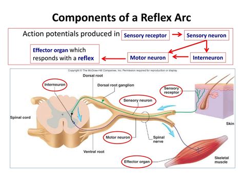
Table of Contents
The Reflex Arc: A Step-by-Step Guide to Understanding Your Body's Automatic Responses
The human body is a marvel of intricate systems, constantly working to maintain balance and respond to its environment. One of the most fascinating aspects of this complex machinery is the reflex arc, a rapid, involuntary response to a stimulus. Understanding the sequence of events in a reflex arc is key to appreciating the nervous system's remarkable efficiency and protective mechanisms. This comprehensive guide will break down each component, placing them in the correct order and exploring their individual roles in this crucial neurological pathway.
What is a Reflex Arc?
A reflex arc is a neural pathway that controls a reflex action. It's a rapid, automatic response to a stimulus that bypasses the brain, allowing for incredibly fast reaction times. This is crucial for protective reflexes, such as withdrawing your hand from a hot stove before you even consciously feel the pain. Imagine the delay if your brain had to process every single sensory input before initiating a response – you’d be in serious trouble! The speed and efficiency of the reflex arc are vital for survival.
This pathway involves specific components working in a precise order. Let's delve into each element and their sequential arrangement:
1. Receptor: Sensing the Stimulus
The journey begins with the receptor, a specialized sensory neuron. Receptors are located throughout the body in various sensory organs and tissues. They are exquisitely sensitive to specific stimuli, such as:
- Mechanoreceptors: Respond to mechanical pressure or distortion, like touch, pressure, vibration, and stretch (found in skin, muscles, and joints).
- Thermoreceptors: Detect changes in temperature (found in skin).
- Nociceptors: Sense painful stimuli, like extreme temperatures, pressure, or chemicals (found throughout the body, but especially in skin and internal organs).
- Photoreceptors: Respond to light (found in the retina of the eye).
- Chemoreceptors: Detect chemicals, like taste and smell (found in taste buds and olfactory epithelium).
When a stimulus (e.g., a sharp pinprick) reaches the receptor, it triggers a change in the receptor's membrane potential. This change is the beginning of the signal transduction process, converting the stimulus into a neural signal. This signal is then transmitted along the sensory neuron. The type of receptor engaged dictates the type of reflex. For instance, a tap on the patellar tendon will activate muscle stretch receptors, leading to the knee-jerk reflex.
2. Sensory Neuron: Transmitting the Signal
Next in the sequence is the sensory neuron, also known as the afferent neuron. This neuron transmits the signal from the receptor to the central nervous system (CNS), which consists of the brain and spinal cord. The sensory neuron's axon carries the nerve impulse towards the CNS. These neurons are typically pseudounipolar, meaning they have a single axon that branches into two, one extending towards the receptor and the other towards the CNS.
The signal transmission across the sensory neuron involves the generation and propagation of action potentials. These are electrical signals that travel along the axon, driven by the movement of ions across the neuron's membrane. The speed of signal transmission depends on factors like the axon's diameter and myelination (the presence of a myelin sheath, which acts as an insulator). Myelinated axons conduct signals much faster than unmyelinated axons.
3. Integration Center: Processing the Information
The integration center is the crucial processing hub within the CNS. This is where the sensory information is received and a decision is made about the appropriate response. In simpler reflexes, this integration takes place within the spinal cord itself, without the brain's direct involvement. This is why reflex actions are so fast. In more complex reflexes, the brain might be involved in processing the information, refining the response, or even overriding the reflex.
The integration center consists of one or more synapses – junctions between neurons. At these synapses, the signal is transmitted from the sensory neuron to an interneuron (in many reflexes, but not all) and then to a motor neuron. Neurotransmitters, chemical messengers, are released at the synapse, enabling communication between neurons. This communication involves the binding of neurotransmitters to receptors on the postsynaptic neuron's membrane, triggering a change in the membrane potential and potentially initiating a new action potential.
4. Motor Neuron: Initiating the Response
Once the integration center has processed the sensory information, the motor neuron, also known as the efferent neuron, takes over. This neuron transmits the signal from the CNS to the effector organ – the muscle or gland that will carry out the response. The motor neuron's axon extends from the CNS to the effector organ, conveying the instructions for action.
Similar to sensory neurons, the signal transmission in motor neurons relies on action potentials. The frequency and pattern of action potentials dictate the strength and duration of the effector organ's response. The motor neuron releases neurotransmitters at the neuromuscular junction (the synapse between the motor neuron and muscle fiber) triggering muscle contraction.
5. Effector: Producing the Response
Finally, the effector carries out the response. The effector is typically a muscle or a gland. In the case of a muscle, the signal from the motor neuron will cause it to contract, leading to a movement. For instance, in the knee-jerk reflex, the effector is the quadriceps muscle, which contracts causing the leg to extend. If the effector is a gland, the signal will stimulate the gland to secrete a substance, such as hormones or enzymes.
The type of response varies depending on the reflex and the effector involved. Reflexes can range from simple muscle contractions to complex glandular secretions. The entire process, from the initial stimulus to the final response, happens in milliseconds, demonstrating the remarkable speed and precision of the reflex arc.
Different Types of Reflex Arcs
While the basic components remain the same, there are variations in the complexity of reflex arcs:
-
Monosynaptic Reflex Arcs: The simplest type, involving only one synapse between the sensory neuron and the motor neuron. The classic knee-jerk reflex is an example. There is no interneuron involved in this type of reflex.
-
Polysynaptic Reflex Arcs: These involve one or more interneurons between the sensory and motor neurons. This allows for more complex processing and coordination of responses, often involving inhibitory interneurons that suppress unwanted muscle activity during a reflex. The withdrawal reflex (pulling your hand away from a hot object) is a polysynaptic reflex, involving several interneurons to coordinate the contraction of flexor muscles and relaxation of extensor muscles in the affected limb. Often reciprocal inhibition is employed to ensure smooth movement.
Clinical Significance of Reflex Arc Testing
Reflex testing is a crucial part of neurological examinations. By assessing the speed, strength, and presence or absence of reflexes, doctors can evaluate the integrity of the nervous system. Abnormalities in reflexes can indicate damage to the nervous system, such as:
-
Hyporeflexia: Diminished or absent reflexes, indicating damage to the sensory or motor neurons or the neuromuscular junction.
-
Hyperreflexia: Exaggerated reflexes, often indicating damage to the upper motor neurons in the brain or spinal cord.
-
Clonus: Rhythmic, involuntary muscle contractions, suggestive of upper motor neuron lesions.
Reflex arc testing helps identify various neurological conditions, such as spinal cord injuries, stroke, multiple sclerosis, and peripheral neuropathies. The precise assessment of reflex responses provides critical information for diagnosis and treatment planning.
Conclusion
The reflex arc is a fundamental aspect of the nervous system, enabling rapid and automatic responses to stimuli. Understanding the sequence of events – receptor, sensory neuron, integration center, motor neuron, and effector – is key to appreciating the intricate workings of this protective mechanism. The speed and precision of reflex actions highlight the remarkable efficiency of the nervous system and their clinical relevance in neurological assessments cannot be overstated. Further exploration into the specific neurotransmitters and ion channels involved in each stage reveals the depth and complexity of this seemingly simple process. The more we understand about the reflex arc, the better we can appreciate the incredible complexity and robustness of the human body.
Latest Posts
Latest Posts
-
Words That End With A S
May 09, 2025
-
25 Is 50 Of What Number
May 09, 2025
-
What Is 9 Percent In Decimal Form
May 09, 2025
-
5 Letter Word Containing S And I
May 09, 2025
-
Difference Between Experimental Probability And Theoretical Probability
May 09, 2025
Related Post
Thank you for visiting our website which covers about Place The Following Parts Of A Reflex Arc In Order. . We hope the information provided has been useful to you. Feel free to contact us if you have any questions or need further assistance. See you next time and don't miss to bookmark.