Picture Of Animal Cell With Labels
Juapaving
Mar 21, 2025 · 7 min read
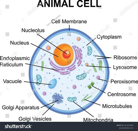
Table of Contents
A Deep Dive into the Animal Cell: Structure, Function, and Stunning Visuals
The animal cell, a fundamental building block of animal life, is a marvel of intricate design. Understanding its structure and the function of its various components is crucial to comprehending the complexities of animal biology. This article will provide a comprehensive exploration of the animal cell, enhanced with detailed descriptions and a labeled diagram to bring this microscopic world to life. We'll explore each organelle, its role, and how these components work together to maintain cellular life.
The Animal Cell: A Microscopic City
Imagine a bustling city, complete with power plants, waste disposal systems, and intricate transportation networks. The animal cell operates in a similar manner, with each organelle playing a specific role in maintaining cellular function and homeostasis. While the exact appearance of an animal cell can vary depending on the organism and cell type, certain key organelles are common across all animal cells. To fully appreciate the complexity, let's delve into the individual components, supported by a visual representation of a typical animal cell with its key structures clearly labeled.
(Insert a high-quality labeled image of an animal cell here. The image should clearly show and label the following: Cell Membrane, Cytoplasm, Nucleus, Nucleolus, Ribosomes, Endoplasmic Reticulum (Rough and Smooth), Golgi Apparatus/Golgi Body, Mitochondria, Lysosomes, Centrosome/Centrioles, Vacuoles.)
Key Components of the Animal Cell: A Detailed Look
Let's explore the main organelles and their functions in detail:
1. Cell Membrane (Plasma Membrane): The Protective Barrier
The cell membrane, also known as the plasma membrane, is the outer boundary of the cell. This selectively permeable membrane acts as a gatekeeper, regulating the passage of substances into and out of the cell. It's composed of a phospholipid bilayer, with embedded proteins that facilitate transport, cell signaling, and cell adhesion. The membrane's fluidity allows for dynamic interactions and adaptations to changing cellular environments. Think of it as the cell's security system, controlling what enters and exits.
2. Cytoplasm: The Cellular Interior
The cytoplasm is the jelly-like substance that fills the cell's interior, excluding the nucleus. It's a complex mixture of water, salts, and various organic molecules. Many cellular processes occur within the cytoplasm, including metabolic reactions and protein synthesis. It provides a medium for the organelles to function and interact, acting as both a support structure and a reaction chamber.
3. Nucleus: The Control Center
The nucleus, the cell's command center, houses the cell's genetic material, DNA (deoxyribonucleic acid). DNA is organized into chromosomes, which contain the instructions for building and maintaining the cell. The nucleus is enclosed by a double membrane called the nuclear envelope, which contains nuclear pores that regulate the passage of molecules between the nucleus and cytoplasm. Think of the nucleus as the city hall, directing all the activities within the cell.
4. Nucleolus: Ribosome Factory
Within the nucleus, you'll find the nucleolus, a dense region responsible for ribosome synthesis. Ribosomes are crucial for protein synthesis, and the nucleolus ensures there's a sufficient supply of these essential cellular components.
5. Ribosomes: Protein Synthesizers
Ribosomes are the protein factories of the cell. They translate the genetic code from mRNA (messenger RNA) into proteins. Ribosomes can be found free in the cytoplasm or attached to the endoplasmic reticulum. These tiny structures are incredibly active, constantly churning out proteins vital for cellular function.
6. Endoplasmic Reticulum (ER): The Manufacturing and Transportation Network
The endoplasmic reticulum (ER) is an extensive network of interconnected membranes extending throughout the cytoplasm. There are two types:
- Rough ER: Studded with ribosomes, the rough ER is involved in protein synthesis and modification. Proteins synthesized on the rough ER are often destined for secretion or incorporation into cell membranes.
- Smooth ER: Lacks ribosomes and plays a crucial role in lipid synthesis, carbohydrate metabolism, and detoxification.
The ER is like the cell's extensive highway system, transporting materials throughout the cell.
7. Golgi Apparatus (Golgi Body): The Packaging and Shipping Center
The Golgi apparatus, or Golgi body, is a stack of flattened, membrane-bound sacs. It receives proteins and lipids from the ER, modifies them, sorts them, and packages them into vesicles for transport to other parts of the cell or for secretion. It's the cell's shipping and receiving department, ensuring that proteins and lipids reach their correct destinations.
8. Mitochondria: The Powerhouses
Mitochondria are the powerhouses of the cell, generating energy in the form of ATP (adenosine triphosphate) through cellular respiration. They have their own DNA and ribosomes, suggesting an endosymbiotic origin. Mitochondria are vital for numerous cellular processes that require energy. Consider them the city's power plants, providing the energy needed for all cellular functions.
9. Lysosomes: The Waste Recycling Centers
Lysosomes are membrane-bound organelles containing digestive enzymes. They break down waste products, cellular debris, and foreign substances. They are essential for maintaining cellular cleanliness and preventing the accumulation of harmful materials. Think of lysosomes as the city's sanitation department, cleaning up waste and recycling materials.
10. Centrosome/Centrioles: The Microtubule Organizers
The centrosome, located near the nucleus, is a microtubule-organizing center. It contains two centrioles, cylindrical structures involved in cell division. They play a crucial role in organizing the microtubules that form the mitotic spindle during cell division.
11. Vacuoles: Storage and Transport
Vacuoles are membrane-bound sacs that store various substances, including water, nutrients, and waste products. Animal cells typically have smaller, more numerous vacuoles compared to plant cells. They act as temporary storage units for a variety of cellular materials.
Understanding the Interconnectedness of Organelles
The organelles within an animal cell don't operate in isolation; they work together in a coordinated manner to maintain cellular function. For example, the ER, Golgi apparatus, and vesicles work together in the secretory pathway, transporting proteins from their synthesis site to their final destination. Mitochondria provide the energy needed for many cellular processes, while lysosomes help maintain cellular cleanliness. This intricate interplay ensures the cell functions efficiently and effectively.
Beyond the Basics: Variations in Animal Cells
It's crucial to remember that the depiction of a typical animal cell is a generalized model. Animal cells exhibit significant diversity in structure and function, depending on their specialized roles within the organism. For instance, muscle cells have abundant mitochondria to provide the energy for contraction, while nerve cells have long, slender extensions to transmit signals. This cellular diversity reflects the remarkable adaptability of animal cells to perform a wide range of functions.
Applications and Further Exploration
Understanding animal cell structure is fundamental to various fields, including medicine, biotechnology, and developmental biology. Researchers utilize this knowledge to understand disease mechanisms, develop new therapies, and manipulate cellular processes for various applications. Further exploration into specific cell types and their unique adaptations will continue to provide valuable insights into the intricate world of animal cells. The study of animal cells remains an active area of research, with new discoveries constantly expanding our understanding of these fundamental units of life. Continued advancements in microscopy techniques and molecular biology will undoubtedly reveal even more about the complexities and intricacies of the animal cell.
Conclusion: The Intriguing World of the Animal Cell
The animal cell, a miniature marvel of biological engineering, is a testament to the elegance and efficiency of life's design. By understanding the structure and function of its various components, we gain a deeper appreciation of the complexities that underpin animal life. This article, complemented by a detailed labeled diagram, has provided a comprehensive overview of the animal cell, its organelles, and their interconnected functions. Further exploration into the specifics of different cell types and their specialized roles will undoubtedly deepen our understanding of this fascinating and vital component of the living world.
Latest Posts
Latest Posts
-
What Is The Lowest Common Multiple Of 4 And 8
Mar 21, 2025
-
The Light Dependent Reactions Occur In The Stroma Of The Chloroplast
Mar 21, 2025
-
What Is The Lowest Common Multiple Of 3 And 5
Mar 21, 2025
-
How Many Weeks Are In 49 Days
Mar 21, 2025
-
Three Examples Of Low Kinetic Energy
Mar 21, 2025
Related Post
Thank you for visiting our website which covers about Picture Of Animal Cell With Labels . We hope the information provided has been useful to you. Feel free to contact us if you have any questions or need further assistance. See you next time and don't miss to bookmark.
