Mitosis In An Onion Root Tip
Juapaving
Mar 21, 2025 · 7 min read
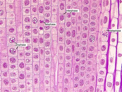
Table of Contents
Mitosis in an Onion Root Tip: A Comprehensive Guide
Mitosis, the process of cell division that results in two identical daughter cells, is fundamental to life. Understanding this process is crucial in biology, and observing it in action provides a valuable hands-on learning experience. The onion root tip, with its actively dividing cells, serves as an excellent model for studying mitosis. This detailed guide will walk you through the process, from preparation to observation and analysis, highlighting the key stages and their significance.
Why Onion Root Tips?
The onion root tip is a popular choice for observing mitosis due to several key advantages:
- High mitotic index: The root tip contains a meristematic region – a zone of rapidly dividing cells – making it easy to find cells undergoing mitosis. This high mitotic index significantly increases the efficiency of observation.
- Ease of access and preparation: Onions are readily available and inexpensive. Preparing the root tip for microscopic observation is relatively straightforward.
- Large, clear cells: The cells in the onion root tip are large enough to be easily observed under a light microscope, and their chromosomes are clearly visible during mitosis.
- Ethical considerations: Unlike animal tissues, using onion root tips raises no ethical concerns.
Preparing the Onion Root Tip for Microscopic Observation
The process of preparing the onion root tip involves several crucial steps, each contributing to the successful visualization of mitotic stages:
1. Growing the Onion Root:
- Select a healthy onion bulb.
- Place the onion in a glass of water, ensuring that the basal part (the bottom) is submerged.
- Allow the onion to grow roots for approximately 5-7 days. The roots should be about 2-3 cm long for optimal results. This growth period allows for sufficient cell division.
2. Fixing the Root Tip:
- Carefully remove the root tip (approximately 1 cm long) using a sharp razor blade or scalpel. Speed and precision are important here to minimize cell damage.
- Immediately transfer the root tip to a fixative solution. A commonly used fixative is glacial acetic acid and ethanol (1:3 ratio). This fixative preserves the cells and prevents further cell division, ensuring the chromosomes remain intact and clearly visible.
3. Hydrolyzing the Root Tip:
- After fixation (at least 30 minutes), the root tip is hydrolyzed using hydrochloric acid. This process softens the cell walls, making the chromosomes more easily accessible for staining. The exact time and concentration of hydrochloric acid will vary depending on the chosen protocol.
4. Staining the Root Tip:
- After hydrolysis, the root tip is stained using a suitable dye, such as aceto-orcein or Feulgen stain. These stains bind to the chromosomes, making them clearly visible under the microscope. Aceto-orcein is a popular choice due to its ease of use and effectiveness. The staining process takes about 5-10 minutes.
5. Squashing and Mounting the Root Tip:
- The stained root tip is then carefully squashed onto a microscope slide using a coverslip. This process spreads the cells, making it easier to observe individual cells undergoing mitosis. Gentle pressure is crucial to avoid damaging the cells.
- Finally, the coverslip is sealed with clear nail polish or mounting medium to prevent drying and preserve the sample for future observation.
Observing Mitosis Under the Microscope
Once the onion root tip is prepared, it's ready for observation under a light microscope. The microscope should be set to high magnification (400x or higher) to clearly visualize the individual cells and their chromosomes.
Identifying the stages of mitosis: The key stages of mitosis can be identified based on the appearance of the chromosomes. These stages include:
1. Prophase:
- Chromatin condensation: The chromatin condenses into visible chromosomes. Each chromosome consists of two identical sister chromatids joined at the centromere.
- Nuclear envelope breakdown: The nuclear membrane disintegrates, and the chromosomes become scattered within the cytoplasm.
- Spindle fiber formation: The spindle fibers, composed of microtubules, begin to form between the centrosomes, which have migrated to opposite poles of the cell.
2. Metaphase:
- Chromosome alignment: The chromosomes align along the metaphase plate, an imaginary plane equidistant from the two poles of the spindle. This alignment ensures that each daughter cell receives one copy of each chromosome.
- Sister chromatid attachment: Each chromosome is attached to spindle fibers from both poles. This attachment is crucial for the separation of sister chromatids in the next phase.
3. Anaphase:
- Sister chromatid separation: The sister chromatids separate at the centromere and move towards opposite poles of the cell. This separation is driven by the shortening of the spindle fibers.
- Chromosome movement: The separated chromatids (now considered individual chromosomes) migrate towards the poles, appearing V-shaped due to the attachment of spindle fibers.
4. Telophase:
- Chromosome arrival at poles: The chromosomes arrive at the opposite poles of the cell.
- Nuclear envelope reformation: A new nuclear envelope forms around each set of chromosomes, creating two distinct nuclei.
- Chromosome decondensation: The chromosomes begin to decondense, becoming less visible.
- Spindle fiber breakdown: The spindle fibers disintegrate.
5. Cytokinesis:
- Cell division: The cytoplasm divides, resulting in the formation of two separate daughter cells, each with an identical set of chromosomes. In plant cells, a cell plate forms between the two daughter cells, eventually developing into a new cell wall.
Analyzing the Results
After observing the onion root tip under the microscope, you can perform various analyses:
- Mitotic index calculation: Calculate the mitotic index by counting the number of cells in mitosis (in all stages) and dividing by the total number of cells observed. The mitotic index provides a quantitative measure of the rate of cell division.
- Stage duration estimation: While challenging without sophisticated equipment, a relative estimate of the duration of each mitotic stage can be attempted based on the number of cells observed in each stage.
- Chromosome counting: Count the number of chromosomes in metaphase cells to determine the diploid chromosome number of the onion.
Potential Challenges and Troubleshooting
Several challenges can arise during the preparation and observation of the onion root tip. These include:
- Over-squashing: Excessive pressure during the squashing process can damage the cells, making it difficult to observe the chromosomes.
- Uneven staining: Inconsistent staining can make it difficult to visualize the chromosomes clearly in all cells.
- Poor microscope focus: Improper focusing of the microscope can hinder observation.
Careful attention to detail during each step of the preparation process can minimize these challenges.
Applications and Significance of Mitosis Study
Studying mitosis in onion root tips is not just a classroom exercise; it holds significant applications in various fields:
- Cancer research: Understanding the regulation of mitosis is crucial in cancer research, as uncontrolled cell division is a hallmark of cancer. Studying mitosis in model organisms helps unravel the mechanisms underlying this uncontrolled growth.
- Plant breeding: Understanding cell division is essential for developing improved plant varieties through selective breeding and genetic engineering.
- Developmental biology: Mitosis is fundamental to the development of multicellular organisms, and its study provides insights into the processes that shape the form and function of living beings.
- Environmental monitoring: The sensitivity of cell division to environmental factors can be used to assess the toxicity of pollutants and other environmental stressors.
Conclusion:
Observing mitosis in an onion root tip provides a practical and accessible method for understanding this fundamental biological process. The readily available materials, relatively simple preparation, and clear visualization of mitotic stages make this a valuable tool for education and research. Through careful preparation and observation, you can gain a comprehensive understanding of cell division, its regulation, and its importance in various biological contexts. By meticulously following the steps outlined above and addressing potential challenges, you can achieve a successful observation experience, enriching your knowledge of this vital cellular process. The seemingly simple onion root tip reveals the complexity and elegance of life at a cellular level, reminding us of the intricate mechanisms that drive growth, development, and life itself.
Latest Posts
Latest Posts
-
150 Is 25 Of What Number
Mar 28, 2025
-
4 1 Rounded To The Nearest Tenth
Mar 28, 2025
-
The Physical Expression Of A Gene Is Known As The
Mar 28, 2025
-
Which Of The Following Is A Homogeneous Mixture
Mar 28, 2025
-
Which Of The Following Is A Coenzyme
Mar 28, 2025
Related Post
Thank you for visiting our website which covers about Mitosis In An Onion Root Tip . We hope the information provided has been useful to you. Feel free to contact us if you have any questions or need further assistance. See you next time and don't miss to bookmark.
