Light Microscope Vs Transmission Electron Microscope
Juapaving
Mar 29, 2025 · 6 min read
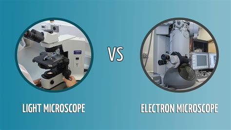
Table of Contents
Light Microscope vs. Transmission Electron Microscope: A Detailed Comparison
The world of microscopy offers two powerful tools for exploring the intricate details of biological specimens and materials: the light microscope and the transmission electron microscope (TEM). While both aim to magnify images beyond the capabilities of the naked eye, they achieve this through vastly different mechanisms, resulting in distinct advantages and limitations. This comprehensive comparison delves into the core principles, applications, and limitations of each technique, providing a clear understanding of when to choose one over the other.
Understanding the Principles: Light vs. Electron Microscopy
The fundamental difference lies in the type of illumination used. Light microscopes utilize visible light to illuminate the specimen, focusing the light rays through a series of lenses to create a magnified image. This is a relatively simple and readily accessible technique.
Transmission electron microscopes (TEMs), on the other hand, employ a beam of electrons instead of light. Electrons have a much shorter wavelength than visible light, allowing for significantly higher resolution. The electrons pass through an ultra-thin specimen, interacting with its structure. The transmitted electrons are then focused by electromagnetic lenses onto a screen or detector, generating a highly magnified image. This approach necessitates complex instrumentation and meticulous sample preparation.
Light Microscopy: A Closer Look
Light microscopy, often referred to as optical microscopy, encompasses various techniques such as bright-field, dark-field, phase-contrast, and fluorescence microscopy. Each technique exploits different properties of light to enhance the visualization of specific features within a sample.
Bright-field microscopy, the most common type, uses transmitted light to illuminate the specimen. The image appears as a result of differences in light absorption by various parts of the sample.
Dark-field microscopy uses scattered light to form the image, resulting in a bright specimen against a dark background. This technique is particularly useful for visualizing transparent specimens.
Phase-contrast microscopy enhances contrast by exploiting differences in refractive index within the specimen. This is vital for observing living cells and unstained specimens.
Fluorescence microscopy uses fluorescent dyes or proteins that emit light at specific wavelengths when excited by light of a shorter wavelength. This technique is exceptionally powerful for visualizing specific molecules or structures within cells.
Transmission Electron Microscopy: A High-Resolution Perspective
TEM offers unmatched resolution, allowing for visualization of structures down to the nanometer scale. This is several orders of magnitude higher than the resolution achievable with light microscopy. The high resolution arises directly from the short wavelength of electrons.
The process involves several key steps:
-
Sample preparation: This is crucial in TEM and involves meticulously preparing ultra-thin sections (typically less than 100 nm thick) of the specimen. This often involves embedding the sample in resin, sectioning it using an ultramicrotome, and staining with heavy metals for contrast.
-
Electron beam illumination: A high-energy electron beam is emitted from an electron gun and accelerated towards the specimen.
-
Electron interaction: The electrons interact with the specimen, some passing through and others being scattered.
-
Image formation: The transmitted electrons are focused by electromagnetic lenses onto a fluorescent screen or a digital detector, producing the image.
The resulting image reveals fine details of the internal structure of cells, tissues, and materials.
Comparative Analysis: Key Differences and Advantages
| Feature | Light Microscope | Transmission Electron Microscope (TEM) |
|---|---|---|
| Resolution | Limited by the wavelength of light (approx. 200 nm) | Extremely high (sub-nanometer resolution possible) |
| Magnification | Typically up to 1500x | Typically up to 1,000,000x |
| Sample Prep | Relatively simple; often requires staining | Complex and time-consuming; requires ultrathin sectioning and staining with heavy metals |
| Cost | Relatively inexpensive | Extremely expensive |
| Specimen | Live or fixed, thick or thin specimens | Requires ultrathin sections; often non-viable |
| Image Type | 2D or 3D (confocal microscopy) | Primarily 2D, though techniques like tomography can provide 3D information |
| Applications | Wide range, including cell biology, histology, microbiology | Material science, nanotechnology, virology, cell ultrastructure |
Advantages of Light Microscopy
- Simplicity and ease of use: Light microscopes are relatively simple to operate and maintain.
- Cost-effectiveness: They are significantly less expensive than TEMs.
- Live cell imaging: Live specimens can be observed, allowing for dynamic processes to be studied in real-time.
- Versatility: Various techniques (bright-field, dark-field, phase-contrast, fluorescence) cater to a broad range of applications.
- Relatively large field of view: Light microscopy allows for a wider field of view than TEM.
Advantages of Transmission Electron Microscopy
- Unmatched resolution: TEM allows visualization of subcellular structures and macromolecular complexes impossible to see with light microscopy.
- High magnification: Achieve magnifications far exceeding those attainable with light microscopy.
- Detailed structural information: Provide intricate details of internal cellular organization and material properties.
- Cryo-TEM: Cryo-electron microscopy allows for imaging of samples in their near-native hydrated state, preserving delicate structures.
Limitations of Light Microscopy
- Lower resolution: Limited ability to resolve fine details at the subcellular level.
- Diffraction limits: The resolution is fundamentally restricted by the wavelength of visible light.
- Specimen preparation can be damaging: Some staining techniques can affect the natural state of the sample.
Limitations of Transmission Electron Microscopy
- High cost: TEMs are expensive to purchase, maintain, and operate.
- Complex sample preparation: Requires extensive training and specialized techniques for sample preparation.
- Vacuum requirement: Samples must be examined under high vacuum, making it unsuitable for live cell imaging.
- Radiation damage: Electron beam can damage the sample.
- Limited field of view: Provides a smaller field of view compared to light microscopy.
Applications: Where Each Technique Excels
Both light and electron microscopy have found widespread applications in various scientific disciplines. The choice of technique depends heavily on the specific research question and the nature of the sample.
Applications of Light Microscopy
- Cell biology: Observing live cells, studying cell division, examining cellular structures like nuclei and organelles.
- Histology: Examining tissue sections, identifying different cell types, diagnosing diseases.
- Microbiology: Identifying bacteria, fungi, and other microorganisms.
- Pathology: Diagnosing diseases by examining tissue samples.
- Materials science: Analyzing the microstructure of certain materials.
Applications of Transmission Electron Microscopy
- Virology: Visualizing virus particles and their interaction with host cells.
- Nanotechnology: Characterizing nanomaterials and their structure.
- Materials science: Analyzing the microstructure of metals, ceramics, and polymers at a very high resolution.
- Cell biology: Examining the ultrastructure of cells, including organelles like mitochondria and ribosomes.
- Medical research: Diagnosing diseases and studying cellular pathology at a high level of detail.
- Cryo-electron microscopy (cryo-TEM): Determining the 3D structure of proteins and macromolecular complexes.
Conclusion: Choosing the Right Microscope
The choice between a light microscope and a transmission electron microscope hinges on the specific research needs. Light microscopy offers a versatile, accessible, and often sufficient tool for many applications, particularly when live cell imaging or a larger field of view is needed. However, for the highest resolution and the detailed visualization of ultrastructure, the TEM remains unparalleled. In some cases, a combined approach, using both techniques, may provide the most complete picture. The researcher must carefully consider the advantages and limitations of each technique to select the most appropriate tool for their investigation. Advances in both light and electron microscopy continue to expand their capabilities, pushing the boundaries of biological and materials science discovery.
Latest Posts
Latest Posts
-
Depreciation Is A Source Of Cash Inflow Because
Mar 31, 2025
-
How To Measure Diameter Of Ball
Mar 31, 2025
-
Molar Mass Of Al Oh 3
Mar 31, 2025
-
An Objects Resistance To A Change In Motion
Mar 31, 2025
-
Why Is The Earth Called A Unique Planet Short Answer
Mar 31, 2025
Related Post
Thank you for visiting our website which covers about Light Microscope Vs Transmission Electron Microscope . We hope the information provided has been useful to you. Feel free to contact us if you have any questions or need further assistance. See you next time and don't miss to bookmark.
