Label The Parts Of A Animal Cell
Juapaving
Mar 21, 2025 · 8 min read
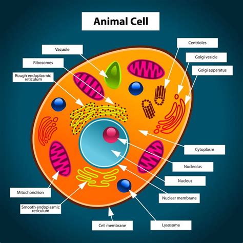
Table of Contents
Labeling the Parts of an Animal Cell: A Comprehensive Guide
Understanding the intricate machinery of a cell is fundamental to grasping the complexities of life itself. Animal cells, the building blocks of animals, are bustling hubs of activity, each component playing a crucial role in maintaining the organism's overall health and function. This comprehensive guide will delve into the detailed structure of an animal cell, providing a clear explanation of each organelle and its vital function. We'll explore their individual roles and how they work together in a coordinated fashion to sustain life. By the end, you'll be able to confidently label the various parts of an animal cell and understand their significance in cellular processes.
The Cell Membrane: The Gatekeeper
Let's start with the outermost layer: the cell membrane, also known as the plasma membrane. This is a selectively permeable barrier, meaning it controls what enters and exits the cell. Think of it as a sophisticated bouncer at an exclusive club, allowing only specific molecules to pass through.
Key Functions of the Cell Membrane:
- Regulation of Transport: The cell membrane carefully regulates the movement of substances like nutrients, waste products, and ions. It achieves this through various mechanisms including passive transport (diffusion, osmosis) and active transport (requiring energy).
- Cell Signaling: Receptor proteins embedded in the membrane receive signals from the environment, triggering cellular responses crucial for growth, division, and interaction with other cells.
- Maintaining Cell Shape: The membrane provides structural support, contributing to the overall shape and integrity of the cell.
- Cell-to-Cell Recognition: Specific molecules on the membrane surface allow cells to recognize and interact with each other, a critical aspect in tissue formation and immune responses.
The cell membrane is primarily composed of a phospholipid bilayer, with hydrophobic tails facing inward and hydrophilic heads facing outward. Embedded within this bilayer are various proteins that carry out the diverse functions mentioned above.
The Cytoplasm: The Cell's Internal Environment
The cytoplasm is the jelly-like substance filling the cell between the cell membrane and the nucleus. It's a dynamic environment where many cellular processes occur. It's not just a passive filler; it's a highly organized space where organelles are suspended and numerous metabolic reactions take place.
Key Functions of the Cytoplasm:
- Site of Metabolic Reactions: Many important biochemical reactions, including glycolysis (the first step in cellular respiration), occur within the cytoplasm.
- Organelle Suspension: The cytoplasm provides a medium for suspending and supporting the various organelles within the cell.
- Cytoplasmic Streaming: The movement of the cytoplasm facilitates the transport of materials within the cell.
The cytoplasm is composed mainly of water, salts, and various organic molecules. It’s a constantly changing environment reflecting the cell's dynamic state.
The Nucleus: The Control Center
The nucleus is the prominent, centrally located organelle in most animal cells. It's often described as the cell's "control center" because it houses the cell's genetic material – deoxyribonucleic acid (DNA). This DNA contains the instructions for building and maintaining the entire organism.
Key Functions of the Nucleus:
- DNA Replication: The nucleus is where DNA replication occurs, ensuring that genetic information is accurately duplicated before cell division.
- Transcription: The process of transcribing DNA into RNA, the messenger molecule carrying genetic instructions to ribosomes, takes place within the nucleus.
- Regulation of Gene Expression: The nucleus controls which genes are expressed (activated) and which are repressed, allowing for precise control over cellular functions.
The nucleus is surrounded by a double membrane called the nuclear envelope, which is perforated by nuclear pores allowing for the selective transport of molecules between the nucleus and the cytoplasm. Inside the nucleus, you'll find the nucleolus, a dense region where ribosomes are assembled.
Ribosomes: Protein Factories
Ribosomes are small, granular organelles responsible for protein synthesis – the process of building proteins from amino acids. These are the cell's protein factories, translating the genetic code from RNA into functional proteins.
Key Functions of Ribosomes:
- Protein Synthesis: Ribosomes bind to messenger RNA (mRNA) and use it as a template to assemble amino acids into polypeptide chains, which fold into functional proteins.
- Location: Ribosomes can be found free-floating in the cytoplasm or attached to the endoplasmic reticulum. Free ribosomes produce proteins used within the cytoplasm, while those attached to the ER synthesize proteins destined for export or for use within the cell membrane.
Ribosomes are composed of ribosomal RNA (rRNA) and proteins. They are essential for all cellular processes, as proteins perform a wide array of functions within the cell and throughout the organism.
Endoplasmic Reticulum (ER): The Manufacturing and Transport System
The endoplasmic reticulum (ER) is a network of interconnected membranes extending throughout the cytoplasm. It comes in two forms: rough ER and smooth ER.
Rough Endoplasmic Reticulum (RER):
The rough ER is studded with ribosomes, giving it its characteristic "rough" appearance. Its primary function is protein synthesis and modification. Proteins synthesized on the RER ribosomes are often destined for secretion from the cell or incorporation into cell membranes.
Smooth Endoplasmic Reticulum (SER):
The smooth ER lacks ribosomes and plays a role in lipid synthesis, detoxification, and calcium storage. It synthesizes lipids like phospholipids and steroids, and it detoxifies harmful substances.
Key Functions of the ER:
- Protein Synthesis and Modification: RER is involved in the synthesis, folding, and modification of proteins.
- Lipid Synthesis: SER synthesizes lipids, including phospholipids and steroids.
- Detoxification: SER plays a role in detoxifying harmful substances.
- Calcium Storage: SER stores calcium ions, which are crucial for various cellular processes.
Golgi Apparatus: The Processing and Packaging Center
The Golgi apparatus (or Golgi complex) is a stack of flattened, membrane-bound sacs called cisternae. It acts as the cell's processing and packaging center, receiving proteins and lipids from the ER and modifying, sorting, and packaging them for transport to their final destinations.
Key Functions of the Golgi Apparatus:
- Protein and Lipid Modification: The Golgi modifies proteins and lipids received from the ER, adding sugar groups or other modifications.
- Sorting and Packaging: The Golgi sorts and packages proteins and lipids into vesicles for transport to other organelles or secretion from the cell.
- Lysosome Formation: The Golgi is involved in the formation of lysosomes.
Lysosomes: The Recycling Centers
Lysosomes are membrane-bound organelles containing digestive enzymes. They act as the cell's recycling centers, breaking down waste products, cellular debris, and ingested materials.
Key Functions of Lysosomes:
- Waste Breakdown: Lysosomes break down waste products, cellular debris, and worn-out organelles.
- Autophagy: Lysosomes participate in autophagy, a process where the cell breaks down and recycles its own components.
- Defense: Lysosomes can engulf and destroy invading pathogens.
Mitochondria: The Powerhouses
Mitochondria are often called the "powerhouses" of the cell because they are the primary site of cellular respiration – the process of converting energy from nutrients into a usable form, ATP (adenosine triphosphate).
Key Functions of Mitochondria:
- ATP Production: Mitochondria generate ATP, the cell's primary energy currency.
- Cellular Respiration: Mitochondria carry out cellular respiration, a process involving several steps to extract energy from glucose.
- Apoptosis Regulation: Mitochondria play a role in regulating programmed cell death (apoptosis).
Vacuoles: Storage and Transport
Vacuoles are membrane-bound sacs used for storage and transport. In animal cells, they are typically smaller and more numerous than in plant cells.
Key Functions of Vacuoles:
- Storage: Vacuoles store various substances, including water, nutrients, and waste products.
- Transport: Vacuoles transport materials within the cell.
Centrosomes and Centrioles: Role in Cell Division
Centrosomes are microtubule-organizing centers found near the nucleus. They contain a pair of centrioles, cylindrical structures involved in cell division.
Key Functions of Centrosomes and Centrioles:
- Microtubule Organization: Centrosomes organize microtubules, which are involved in cell structure and movement.
- Cell Division: Centrioles play a crucial role in cell division, forming the mitotic spindle that separates chromosomes during mitosis.
Peroxisomes: Detoxification and Lipid Metabolism
Peroxisomes are small, membrane-bound organelles involved in various metabolic processes, including detoxification and lipid metabolism.
Key Functions of Peroxisomes:
- Detoxification: Peroxisomes break down harmful substances, such as hydrogen peroxide.
- Lipid Metabolism: Peroxisomes play a role in the breakdown of fatty acids and other lipids.
Cytoskeleton: Cellular Framework
The cytoskeleton is a network of protein filaments extending throughout the cytoplasm. It provides structural support, maintains cell shape, and facilitates cell movement.
Key Functions of the Cytoskeleton:
- Structural Support: The cytoskeleton provides structural support to the cell.
- Cell Shape Maintenance: It helps maintain the cell's shape and organization.
- Cell Movement: The cytoskeleton is involved in various types of cell movement, including cell division and cytoplasmic streaming.
This detailed overview provides a comprehensive understanding of the various parts of an animal cell and their respective functions. Remember, these organelles work together in a highly coordinated manner to maintain the cell's integrity and carry out the essential processes of life. Understanding these components is critical for appreciating the wonders of biology and the complexity of living organisms. Further exploration into the intricate biochemical pathways and interactions within the cell will reveal even greater depths of this fascinating subject.
Latest Posts
Latest Posts
-
What Is The Lowest Common Multiple Of 4 And 8
Mar 21, 2025
-
The Light Dependent Reactions Occur In The Stroma Of The Chloroplast
Mar 21, 2025
-
What Is The Lowest Common Multiple Of 3 And 5
Mar 21, 2025
-
How Many Weeks Are In 49 Days
Mar 21, 2025
-
Three Examples Of Low Kinetic Energy
Mar 21, 2025
Related Post
Thank you for visiting our website which covers about Label The Parts Of A Animal Cell . We hope the information provided has been useful to you. Feel free to contact us if you have any questions or need further assistance. See you next time and don't miss to bookmark.
