Identify The Stages Of Meiosis On The Diagram
Juapaving
Mar 21, 2025 · 6 min read
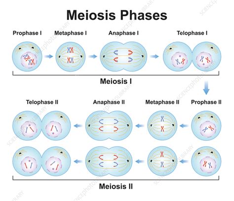
Table of Contents
Identifying the Stages of Meiosis on a Diagram: A Comprehensive Guide
Meiosis, the specialized type of cell division that produces gametes (sex cells), is a complex process involving two successive divisions: Meiosis I and Meiosis II. Understanding the distinct stages of each division is crucial for grasping the fundamentals of genetics and reproduction. This comprehensive guide will equip you with the knowledge to accurately identify the stages of meiosis depicted in any diagram, focusing on the key morphological changes within the cell.
Meiosis I: Reducing the Chromosome Number
Meiosis I is the reductional division, where the homologous chromosomes are separated, reducing the chromosome number from diploid (2n) to haploid (n). This ensures that when gametes fuse during fertilization, the resulting zygote maintains the correct diploid number of chromosomes. Let's break down the stages:
1. Prophase I: A Lengthy and Complex Stage
Prophase I is the longest and most intricate phase of meiosis I. Several key events occur:
- Leptotene: Chromosomes begin to condense, becoming visible under a microscope as long, thin threads. Each chromosome consists of two sister chromatids joined at the centromere, though the sister chromatids are often indistinguishable at this stage.
- Zygotene: Homologous chromosomes (one from each parent) begin to pair up, a process called synapsis. The paired homologous chromosomes are referred to as bivalents. A protein structure called the synaptonemal complex forms between the homologous chromosomes, holding them together tightly.
- Pachytene: The synaptonemal complex is fully formed, and crossing over occurs. Crossing over is the exchange of genetic material between non-sister chromatids of homologous chromosomes. This crucial process shuffles alleles, increasing genetic diversity. The points where crossing over occurs are visible as chiasmata (singular: chiasma).
- Diplotene: The synaptonemal complex disassembles, and homologous chromosomes begin to separate. However, they remain connected at the chiasmata, which are visible as cross-shaped structures.
- Diakinesis: Chromosomes continue to condense and shorten. The nuclear envelope breaks down, and the spindle fibers begin to form. The chiasmata move towards the ends of the chromosomes, a process known as terminalization.
Identifying Prophase I in a Diagram: Look for paired homologous chromosomes (bivalents), the presence of chiasmata, condensed chromosomes, and the eventual breakdown of the nuclear envelope. The specific sub-stages within Prophase I (Leptotene, Zygotene, Pachytene, Diplotene, Diakinesis) can be differentiated by the degree of chromosome condensation, the presence and structure of the synaptonemal complex, and the location of the chiasmata.
2. Metaphase I: Alignment on the Metaphase Plate
In Metaphase I, the paired homologous chromosomes align along the metaphase plate (the equator of the cell). Each homologous chromosome is attached to a spindle fiber from opposite poles of the cell. This arrangement ensures that each daughter cell will receive one chromosome from each homologous pair.
Identifying Metaphase I in a Diagram: Look for homologous chromosome pairs aligned at the metaphase plate, attached to spindle fibers emanating from opposite poles. The chromosomes are highly condensed.
3. Anaphase I: Separation of Homologous Chromosomes
During Anaphase I, the homologous chromosomes are separated and pulled towards opposite poles of the cell by the spindle fibers. Sister chromatids remain attached at the centromere. This is a key difference between Anaphase I and Anaphase II.
Identifying Anaphase I in a Diagram: Observe the separation of homologous chromosomes, with each chromosome moving towards opposite poles. Sister chromatids remain connected.
4. Telophase I and Cytokinesis: Two Haploid Cells
In Telophase I, the chromosomes arrive at the poles of the cell. The nuclear envelope may reform, and the chromosomes may decondense slightly. Cytokinesis, the division of the cytoplasm, follows Telophase I, resulting in two haploid daughter cells. Each daughter cell contains one chromosome from each homologous pair, but each chromosome still consists of two sister chromatids.
Identifying Telophase I and Cytokinesis in a Diagram: Look for the arrival of chromosomes at opposite poles, potential reformation of the nuclear envelope, chromosome decondensation, and the formation of a cleavage furrow (in animal cells) or cell plate (in plant cells) indicating cytokinesis.
Meiosis II: Separating Sister Chromatids
Meiosis II is similar to mitosis in that it involves the separation of sister chromatids. However, it starts with haploid cells, resulting in four haploid daughter cells, each with a single copy of each chromosome.
1. Prophase II: Chromosomes Condense Again
Prophase II is much shorter than Prophase I. The chromosomes condense again, and the nuclear envelope breaks down (if it reformed during Telophase I). The spindle fibers begin to form.
Identifying Prophase II in a Diagram: Observe the recondensation of chromosomes, breakdown of the nuclear envelope (if present), and the formation of the spindle apparatus.
2. Metaphase II: Chromosomes Align at the Metaphase Plate
In Metaphase II, the individual chromosomes (each consisting of two sister chromatids) align along the metaphase plate. Each sister chromatid is attached to a spindle fiber from opposite poles.
Identifying Metaphase II in a Diagram: Look for individual chromosomes aligned at the metaphase plate, attached to spindle fibers from opposite poles.
3. Anaphase II: Separation of Sister Chromatids
During Anaphase II, the sister chromatids are finally separated and pulled towards opposite poles of the cell by the spindle fibers.
Identifying Anaphase II in a Diagram: Observe the separation of sister chromatids, with each chromatid (now a chromosome) moving towards opposite poles.
4. Telophase II and Cytokinesis: Four Haploid Cells
In Telophase II, the chromosomes arrive at the poles of the cell. The nuclear envelope reforms, and the chromosomes decondense. Cytokinesis follows Telophase II, resulting in four haploid daughter cells, each genetically unique due to crossing over and independent assortment.
Identifying Telophase II and Cytokinesis in a Diagram: Look for the arrival of chromosomes at opposite poles, reformation of the nuclear envelope, chromosome decondensation, and the completion of cytokinesis, resulting in four separate haploid daughter cells.
Key Differences Between Meiosis I and Meiosis II
| Feature | Meiosis I | Meiosis II |
|---|---|---|
| Purpose | Reductional division (2n to n) | Equational division (n to n) |
| Homologous Chromosomes | Separate | Remain separate |
| Sister Chromatids | Remain attached during Anaphase I | Separate during Anaphase II |
| Crossing Over | Occurs in Prophase I | Does not occur |
| Genetic Variation | High, due to crossing over and independent assortment | No further increase in genetic variation |
Practical Tips for Identifying Meiosis Stages
- Chromosome Condensation: Pay close attention to the degree of chromosome condensation. Chromosomes are highly condensed in Metaphase I and II.
- Nuclear Envelope: The presence or absence of the nuclear envelope is a key indicator. It typically breaks down in Prophase and reforms in Telophase.
- Spindle Fibers: The presence and arrangement of spindle fibers indicate the stage of division.
- Chromosome Alignment: The arrangement of chromosomes at the metaphase plate is distinct in Metaphase I (homologous pairs) and Metaphase II (individual chromosomes).
- Sister Chromatid Separation: The separation of sister chromatids occurs only in Anaphase II.
By carefully observing these morphological characteristics within a diagram, you can confidently identify the different stages of meiosis. Remember to consider the context of the cell type and the overall process to make accurate identifications. This detailed guide should serve as a valuable resource for students and anyone interested in understanding the intricacies of this fundamental biological process. Regular practice with diagrams will solidify your understanding and enable you to quickly and accurately identify each stage of meiosis. Mastering the identification of these stages is crucial for a thorough understanding of genetics and its implications.
Latest Posts
Latest Posts
-
What Is Sulphur Used For In Everyday Life
Mar 27, 2025
-
Solid Has A Definite Shape And Volume
Mar 27, 2025
-
What Is The Percentage Of 32 Out Of 40
Mar 27, 2025
-
Words With Ie In Them 5 Letters
Mar 27, 2025
-
What Are The Pros Of Fossil Fuels
Mar 27, 2025
Related Post
Thank you for visiting our website which covers about Identify The Stages Of Meiosis On The Diagram . We hope the information provided has been useful to you. Feel free to contact us if you have any questions or need further assistance. See you next time and don't miss to bookmark.
