Diff Between Light And Electron Microscope
Juapaving
Apr 07, 2025 · 5 min read
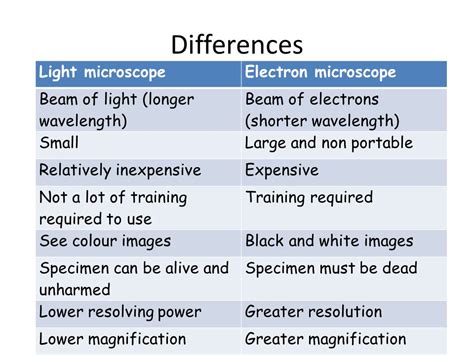
Table of Contents
- Diff Between Light And Electron Microscope
- Table of Contents
- Delving Deep: The Key Differences Between Light and Electron Microscopes
- Understanding Magnification and Resolution: The Cornerstones of Microscopy
- The Light Microscope: A Window to the Microscopic World
- Principles of Operation:
- Types of Light Microscopy:
- Limitations of Light Microscopy:
- The Electron Microscope: Peering into the Nano-World
- Principles of Operation:
- Types of Electron Microscopy:
- Advantages of Electron Microscopy:
- Limitations of Electron Microscopy:
- A Direct Comparison: Light vs. Electron Microscopy
- Conclusion: Choosing the Right Microscope
- Latest Posts
- Latest Posts
- Related Post
Delving Deep: The Key Differences Between Light and Electron Microscopes
The world is teeming with life, structures, and materials far too small for the naked eye to perceive. To explore this microscopic universe, scientists rely on powerful tools: microscopes. While both light and electron microscopes magnify images, their underlying principles, capabilities, and applications differ significantly. This comprehensive guide delves into the core distinctions between these two crucial scientific instruments, highlighting their strengths and limitations.
Understanding Magnification and Resolution: The Cornerstones of Microscopy
Before diving into the specifics of light and electron microscopes, let's clarify two fundamental concepts: magnification and resolution.
-
Magnification: This refers to the ability of a microscope to enlarge an image. A higher magnification means a larger, more detailed view of the specimen.
-
Resolution: This signifies the microscope's capacity to distinguish between two closely positioned objects. High resolution means the ability to see finer details and separate structures that are very close together. Resolution is arguably more crucial than magnification; a highly magnified image with poor resolution is blurry and uninformative.
The Light Microscope: A Window to the Microscopic World
The light microscope, or optical microscope, uses visible light and a system of lenses to magnify specimens. Its simplicity and relative affordability have made it a staple in numerous scientific disciplines for centuries.
Principles of Operation:
Light microscopes work by passing light through a specimen, which is then magnified by a series of lenses. The objective lens, positioned closest to the specimen, creates a magnified real image. This image is further magnified by the eyepiece lens, resulting in a virtual image that the observer sees.
Types of Light Microscopy:
Several variations of light microscopy exist, each optimized for specific applications:
-
Bright-field microscopy: This is the most common type, using transmitted light to illuminate the specimen. Stained specimens are generally needed for optimal contrast.
-
Dark-field microscopy: This technique illuminates the specimen from the side, resulting in a bright specimen against a dark background. It's particularly useful for observing unstained, transparent specimens.
-
Phase-contrast microscopy: This method enhances contrast in transparent specimens by exploiting differences in the refractive index of various structures. It's ideal for observing living cells without staining.
-
Fluorescence microscopy: This technique uses fluorescent dyes or proteins to label specific structures within the specimen. Excitation light causes the labeled structures to emit light at a longer wavelength, providing high contrast and specificity.
-
Confocal microscopy: A sophisticated variation of fluorescence microscopy, confocal microscopy uses a pinhole to eliminate out-of-focus light, resulting in sharper, three-dimensional images.
Limitations of Light Microscopy:
Despite its versatility, the light microscope has limitations:
-
Resolution limit: Due to the diffraction of light, the resolving power of a light microscope is limited to approximately 200 nanometers. This means that structures smaller than this cannot be clearly resolved.
-
Specimen preparation: Depending on the microscopy technique, specimen preparation can be time-consuming and may introduce artifacts. Certain techniques require staining, which can kill living cells.
The Electron Microscope: Peering into the Nano-World
The electron microscope utilizes a beam of electrons instead of light to illuminate the specimen. The significantly shorter wavelength of electrons allows for far higher resolution than light microscopy, enabling visualization of structures at the nanometer scale.
Principles of Operation:
Electron microscopes use electromagnetic lenses to focus a beam of electrons onto the specimen. The interaction of the electrons with the specimen generates an image, which is then captured and displayed.
Types of Electron Microscopy:
Two primary types of electron microscopy exist:
-
Transmission Electron Microscopy (TEM): In TEM, a beam of electrons is transmitted through a very thin specimen. The electrons that pass through are detected, creating an image based on the electron density of different structures. TEM provides high resolution, allowing visualization of internal cellular structures and even individual molecules.
-
Scanning Electron Microscopy (SEM): SEM scans the surface of a specimen with a focused electron beam. The scattered electrons are detected, generating a three-dimensional image of the specimen's surface. SEM is excellent for visualizing surface features and topography.
Advantages of Electron Microscopy:
Electron microscopy offers several key advantages:
-
High resolution: The significantly shorter wavelength of electrons enables much higher resolution than light microscopy, revealing fine details impossible to see with light.
-
Detailed structural information: Electron microscopy provides detailed structural information about cells, tissues, and materials at the nanometer scale.
-
Versatility: Both TEM and SEM offer distinct advantages, catering to diverse research needs.
Limitations of Electron Microscopy:
Electron microscopy is not without limitations:
-
Sample preparation: Specimen preparation for electron microscopy is often complex, time-consuming, and can introduce artifacts. Samples generally need to be dehydrated and sometimes coated with a conductive material.
-
Cost and maintenance: Electron microscopes are expensive to purchase and maintain, requiring specialized expertise for operation.
-
Vacuum environment: Electron microscopy requires a high vacuum environment, which can be incompatible with certain specimens, particularly living cells.
-
Radiation damage: The electron beam can damage the specimen, particularly biological samples, limiting observation time.
A Direct Comparison: Light vs. Electron Microscopy
| Feature | Light Microscope | Electron Microscope |
|---|---|---|
| Resolution | ~200 nm | < 0.1 nm (TEM), ~1 nm (SEM) |
| Magnification | Up to 1500x | Up to 1,000,000x (TEM), up to 300,000x (SEM) |
| Sample Prep | Relatively simple, often requires staining | Complex, often involves dehydration and coating |
| Cost | Relatively inexpensive | Very expensive |
| Maintenance | Relatively low | High |
| Specimen Type | Live or fixed samples, thicker specimens | Typically fixed, thin sections (TEM), solid (SEM) |
| Imaging Mode | Light transmission/reflection | Electron transmission/scattering |
| Environment | Ambient | High vacuum |
| Applications | Cell biology, histology, microbiology | Materials science, nanotechnology, cell biology |
Conclusion: Choosing the Right Microscope
The choice between a light and electron microscope depends heavily on the specific research question and the nature of the specimen. Light microscopy is well-suited for observing larger structures and living cells, offering a balance of simplicity, affordability, and versatility. Electron microscopy, however, provides unparalleled resolution, revealing the intricate details of cellular structures and materials at the nanoscale. While it demands significant investment and expertise, its power to visualize the ultrastructure is indispensable for numerous scientific advancements. In many cases, researchers utilize both types of microscopy to obtain a comprehensive understanding of their samples, leveraging the strengths of each technique.
Latest Posts
Latest Posts
-
Finding The Area Between Two Curves Calculator
Apr 11, 2025
-
Lipopolysaccharide Is An Important Cell Wall Component Of
Apr 11, 2025
-
Formula Sheet For Area And Perimeter
Apr 11, 2025
-
Derivative Of Harmonic Function Is Harmonic
Apr 11, 2025
-
Table Of Square And Cube Roots
Apr 11, 2025
Related Post
Thank you for visiting our website which covers about Diff Between Light And Electron Microscope . We hope the information provided has been useful to you. Feel free to contact us if you have any questions or need further assistance. See you next time and don't miss to bookmark.